Introduction
One of the greatest assets a person can have is a smile that shows beautiful, natural teeth. Esthetics is a prime consideration in today’s population, with an ever increasing demand for a sparkling smile. In fact today’s patient pays more attention to cosmetics than ever before & teeth are a key consideration in personal appearance. In today’s modern civilized cosmetic world, well contoured & well aligned white teeth set the standard for beauty.
There are many methods to improve esthetics like bleaching, composite restorations, veneers, crowns, etc. Today’s esthetic dentistry seeks to improve the appearance of patients’ smiles with a minimally invasive approach. Tooth discoloration is one reason patients seek esthetic dental treatment. The etiology of dental discoloration is multifactorial.[1]
Discolorations are classified as being extrinsic (attached to the surface of the tooth) or intrinsic (incorporated into the tooth matrix). Although extrinsic discoloration can be removed with a prophylactic cleaning procedure, intrinsic staining necessitates chemical bleaching.[1]
Three treatments are currently popular for removing a variety of intrinsic stains from vital teeth. These are enamel microabrasion technique[2],[3],[4], office bleaching with hydrogen peroxide[5], [6], [7], [8], [9] & home bleaching with carbamide peroxided[10]. While each technique with its accompanying chemical agent has its place in an armamentarium of vital tooth stain removers, none meets the needs of all stains & patient situations.[11] Macroabrasion is another technique which is not followed routinely.
It has been suggested that an approach combining various techniques of bleaching can compensate for their individual shortcomings & extend the effectiveness of this conservative approach to care[12],[13],[14],[15]. A combined technique can result in the successful treatment of some stains where previously only masking procedures with resin bonding, veneers & crowns were effective.[11]
Techniques
Macroabrasion or Megabrasion:
Some enamel defects or white spots on teeth that do not respond to microabrasion and bleaching may respond better to macroabrasion. It is an alternative technique for the removal of localized, superficial white spots & other stains or defects. The discolored enamel contains an increased amount of organic matrix, which is not an adequate substrate for adhesion of dental materials. When the lesion is generally restricted to enamel, its elimination will not result in exposure of dentin (Magne 1997).[16]
Macroabrasion involves removing the outer layer of opaque enamel using a 12-fluted composite finishing bur or a fine grit finishing diamond in a high-speed handpiece with water-air spray. Light, intermittent pressure is used with careful monitoring of removal of tooth structure so as to avoid irreversible damage to the tooth.[17]
After removal of the defect or on termination of any further removal of tooth structure, a 30-fluted, composite finishing bur is used to remove any facets or striations created by the previous instruments. Final polishing is accomplished with an abrasive rubber point.
This technique is advantageous in being considerably faster. Also it does not require the use of a rubber dam or special instrumentation. Gross removal of the defect can be easily accomplished with macroabrasion. High-speed instrumentation as used in macroabrasion is technique sensitive and can have catastrophic results if the clinician fails to use extreme caution. It also makes way for the active reagents of bleaching system to penetrate.
Office bleaching:
Office bleaching techniques rely on the use of 30 to 35% preparations of hydrogen peroxide. Such techniques have been used to remove stains and discolorations that reside in both enamel and the dentin.[11] Its mechanism relies on an oxidation process to reduce large, pigmented molecules to smaller, less noticeable ones[5]. Hydrogen peroxide is a caustic agent. The soft tissue may be protected, usually with a rubber dam. Hydrogen peroxide is capable of penetrating through the tooth to the pulp but with no long-term pulpal reaction[18]. It is suitable for intrinsic stains that are either developmental or acquired in origin.[11]
Hydrogen peroxide is applicable to stains that reside deep in enamel and dentin as well as the superficial enamel. The techniques traditionally associated with office hydrogen peroxide have required several office visits. Nowadays gingival isolation is done via a Top Dam and a 30 to 35% hydrogen peroxide solution is applied to the teeth in liquid or gel form for about 15 minutes cycle.
Another approach depends on activation of the hydrogen peroxide using a composite resin curing light. At the end of the bleaching session the bleach is rinsed from the teeth. The patient repeats the process, usually within 7 to 10 days if required.
Office bleaching techniques can successfully remove many stains that acid abrasion can. They have an advantage of removing deep enamel & dentin retained stains without the loss of tooth structure. It has several disadvantages too. It usually requires several office visits, with consequent increases in overall cost. Additionally, the results of this approach are often unpredictable with relapse and retreatment a common complaint of dentists and patients alike. It does not remove white discolorations, although it can lessen the appearance of whiteness in the stains by lightening adjacent tooth structure. This office bleaching can provide a solution in case there is a lack of compliance with home bleaching.
Following are the reports of few cases being presented to demonstrate how more than one chemical agent or technique can meet the needs of individual stains & patient situations.
Case Reports
Patient 1
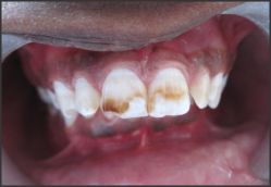
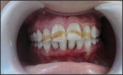
A 20 year old female patient was referred to the Department of Conservative Dentistry & Endodontics of S.G.T. Dental College, Hospital & Research Institute, with a chief complaint of presence of localized stains confined to permanent maxillary incisors (Figure 1).
Clinical examination revealed brown bands on maxillary central incisors with yellowish brown discolorations on lateral incisors.
These brown stains were diagnosed to be due to Fluorosis (Grade 3), as patient was a resident of Haryana, where fluoride levels of water are high.
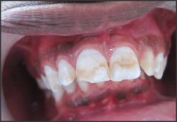
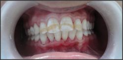
The treatment plan was to use a combination therapy of macroabrasion & bleaching. Macroabrasion was done to remove these typically superficial discolorations using mild abrasion of surface enamel using a fine composite finishing diamond bur, once complete oral prophylaxis had been done (Figure 2).
The brown stains however, were not completely removed after the mild macroabrasion of surface enamel. The residual stain was judged to be seated deep in dentin. To further improve the appearance of these teeth, office bleaching with 35% hydrogen peroxide containing bleaching system (Whiteness HP, FGM, SC, Brazil) (Figure 3) was adopted to lighten the tooth structure adjacent to the white areas so that they would be less noticeable.
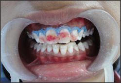
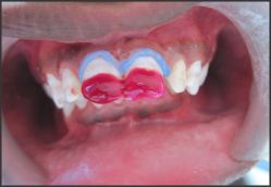
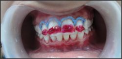
Then with the marginal gingival barrier, the same 35% hydrogen peroxide containing gel was applied on the residual stains of maxillary incisors, according to the manufacturer’s directions, to obtain maximum stain removal (Figure 4). Three applications were done according to the manufacturer’s instructions each of 15 minutes.
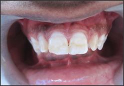
Additional 2 cycles of light curing of 10 seconds each was done for each tooth. (Figure 5)
To allow complete stain removal & enhance the esthetics, a second appointment was given to the patient & office bleaching was again carried out. And the results came out to be appreciable (Figure 6).
Patient 2
A 17 year old male patient, hailing from Gurgaon, Haryana, was referred to the Department of Conservative Dentistry &
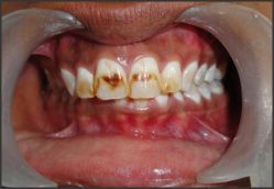 | Figure 1. Pretreatment view where brown stains were localized predominantly on the facial surface of these teeth.
 |
 | Figure 2. Maxillary incisors after macroabrasion to remove superficial brown stains.
|
 | Figure 3. Whiteness HP, FGM (SC, Brazil)
|
 | Figure 4. Operative view of bleaching of maxillary incisors with Whiteness HP, FGM, with a marginal gingival barrier.
|
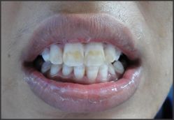 | Figure 5. Teeth immediately after the treatment.
|
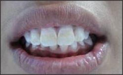 | Figure 6. Maxillary incisors after office bleaching.
|
 | Figure 7. Pretreatment view of brown discoloration predominating the facial aspect of maxillary incisors.
|
 | Figure 8. Maxillary central incisors after macroabrasion to remove superficial brown stains.
|
 | Figure 9. Office bleaching.
|
 | Figure 10. Maxillary central incisors after Office bleaching.
|
Endodontics, of S.G.T Dental College, for his complaint of brown discolored teeth since childhood.
The clinical examination revealed presence of brown discolorations on maxillary central incisors. These brown stains were diagnosed to be due to Fluorosis (Grade 3), as patient was a resident of Haryana, where fluoride levels of water are high (Figure 7).
The treatment plan was to use fine finishing diamond bur to mildly abrade the surface enamel, so as to remove the brown stains & as much of the white discoloration as possible from the superficial enamel of maxillary central incisors.
Note the brown stains have been lightened to light brown on maxillary central incisors in (Figure 8).
For further whitening of teeth, with the marginal gingival barrier, the same 35% hydrogen peroxide containing gel was applied on the residual stains of maxillary incisors, according to the manufacturer’s directions, to obtain maximum stain removal. (Figure 9) shows the operative view of bleaching of maxillary incisors with Whiteness HP, FGM (SC, Brazil) with a marginal gingival barrier.
Three applications were done according to the manufacturer’s instructions each of 15 minutes with additional 2 cycles of light curing of 10 seconds each for each tooth. After the above procedure, brown stains on maxillary central incisors faded away & a better esthetic smile was achieved (Figure 10).
Patient 3
The third patient was a 21 year old female, a resident of Haryana, complaining of presence of localized brown bands on upper front teeth (Figure 11). After the clinical examination, it was concluded that the root cause of the brown discoloration of maxillary incisors was due to Fluorosis.
A combination therapy was again planned for the patient. With a fine finishing diamond bur, dark stains from surface enamel were abraded so as to lighten the stains as seen in the (Figure 12).
Next procedure undertaken was office bleaching with Whiteness HP, FGM, containing 35% Hydrogen Peroxide, under marginal gingival barrier as described in previous cases. (Figure 13)
Esthetic smile was achieved after the procedure (Figure 14).
 | Figure 11. Pretreatment view of discolored maxillary incisors with brown bands.
|
 | Figure 12. Maxillary incisors after macroabrasion.
|
 | Figure 13. Operative view
|
 | Figure 14. Post-operative view of the patient.
|
Discussion
The use of hydrogen peroxide to whiten teeth via an in-office bleaching technique was first reported in 1877. In 1918, Abbot introduced the chairside bleaching method, as it is known today, using 35% hydrogen peroxide with heat & light to boost the oxidation reaction.[1] Even though hydrogen peroxide has been successfully used in dentistry for many years, the mechanism by which bleaching occurs is currently not clearly understood. Several reactions may account for bleaching efficiency, depending on the environmental conditions, such as temperature, pH, ultraviolet light and the presence of some ions. Under alkaline conditions, hydrogen peroxide can undergo an ionic dissociation to give rise to the formation of the perhydroxyl anion. The perhydroxyl anion (HO2-) can be, by itself, the active species in the bleaching process but can also be an electron donor to initiate the formation of free radicals [19]. In addition to the anionic dissociation, hydrogen peroxide can undergo a homolytic cleavage. This reaction is mainly promoted by high temperature and UV light & can result in the appearance of a powerful oxidant agent called hydroxyl radical (HO.) [19]. These free radicals are highly unstable and are therefore, strong oxidative agents which disintegrate the larger chromophores. It results in a shift of the visible absorption spectrum of the compound from a longer to a shorter wavelength, leading to the production of less chromatogenic compounds [20].
Some enamel defects or white spots/opacities on teeth that don’t respond to microabrasion & bleaching may respond better to macroabrasion.
Benbachir N. et al (2007) gave a report illustrated with clinical cases citing the limitations of microabrasion in patients with discolored anterior teeth. In cases with severe enamel defects, megabrasion combined with a minimally invasive adhesive resin composite restoration may present a valuable alternative to microabrasion[21]. Amarlal D. et al (2006) also compiled case reports with relevant clinical photographs of discolored teeth where the treatment regimen included macroabrasion [22].
Bodden MK et al (2003) treated a patient with a level 3 endemic dental fluorosis was treated sequentially by macroabrasion techniques and nightguard vital bleaching. This conservative treatment regimen produced results that were termed "excellent" by the patient and met the goals of the dentists[23].
Macroabrasion removes superficial stains from surface enamel and makes way for the active reagents of bleaching system to penetrate.
Heymann preferred in-office bleaching over home applied technique of bleaching. Although it uses very caustic chemicals it is totally under the dentist’s control, the soft tissue is generally protected from the process and it has the potential for bleaching teeth more rapidly[17].
Conclusion
While macroabrasion, office bleaching, home bleaching and veneering techniques can each successfully solve the problems of many stained & discolored teeth, using the techniques together can help solve many more. Matching the stain with the correct technique requires knowledge of the various intrinsic stains & their potential for stain removal with each chemical agent.
Where stain removal is incomplete with one treatment technique or where multiple stains exist, it is possible to sequence together various techniques for a more efficient & successful result. This combination therapy of macroabrasion and in-office bleaching, for removing intrinsic stains demonstrated effective stain removal and can be considered for most of the tooth discoloration cases and can help in enhancing esthetics.
References
1. Nacer Benbachir, Stefano Ardu, Ivo Krejci. Stectrophotometric evaluation of the efficacy of a new in-office bleaching technique. Quintessence International 2008; 39(4): 299-306.
2. McCloskey RJ. A technique for removal of fluorosis stains. J Am Dent Assoc 1984; 109: 63-64.
3. Croll TP, Cavanaugh RR. Enamel color modifications by controlled hydrochloric acid-pumice abrasions. I: Techniques and examples. Quintessence Int 1986; 17: 81-87.
4. Killian CM, Croll TP. Enamel microabrasion to improve enamel surface texture. J Esthet Dent 1990; 2: 125-128.
5. Bailey R, Christen A. Bleaching of vital teeth with endemic fluorosis. Oral Surg 1968; 26: 871-878.
6. Murrin JR, Barkmeier WM. Chemical treatment of vital teeth with intrinsic stain. Quintessence Int 1982; 13: 6-10.
7. Jordon RE, Boksman L. Conservative vital bleaching treatment of discolored dentition. Compend Contin Educ Dent 1984; 5: 803-808.
8. Feinman RA, Goldstein RE, Garber DA. Bleaching teeth. Chicago: Quintessence. 1987.
9. Feinman RA, Madray G, Yarborough D. Chemical, optical and physiologic mechanisms of bleaching products: a review. Pract Periodont Aesthetic Dent 1991; 3: 32-36.
10. Haywood VB, Heyman HO. Nightguard vital bleaching. Quintessence Int 1989; 20: 173-176.
11. Susan A. McEvoy. Removing intrinsic stains from vital teeth by microabrasion and bleaching. Journal of Esthetic Dentistry, 1995; 7(3): 104-109.
12. McEvoy SA. Chemical agents for removing intrinsic stains from vital teeth. II: Current techniques and their clinical application. Quintessence Int 1989; 20: 379-384.
13. Croll TP. Enamel microabrasion followed by dental bleaching. Case reports. J Esthet Dent 1992; 23: 317-321.
14. Cvitko E, Swift EJ, Denehy GE. Improved esthetics with a combined bleaching technique: a case report. Quintessence Int 1992; 23: 91-93.
15. Killian CM. Conservative color improvement for teeth with fluorosis type stain. J Am Dent Assoc 1993; 124: 72-74.
16. Bleaching techniques in Restorative Dentistry by Linda Greenwall. Pg 207-208.
17. Harald O. Heymann. Sturdevant’s Art & Science of Operative Dentistry. Fifth edition, Mosby, Elsevier; 2006
18. Bowles WH, Ugwuneri Z. Pulp chamber penetration by hydrogen peroxide following vital bleaching procedures. J Endod 1987; 8: 375-377.
19. Maryline Minoux, Rene Serfaty. Vital tooth bleaching: Biologic adverse effects—A review. Quintessence Int. 2008; 39(8): 645-59.
20. Seghi RR, Denry I. Effects of external bleaching on indentation and abrasion characteristics of human enamel in vitro. J Dent Res 1992; 71: 1340-1344.
21. Benbachir N, Ardu S, Krejci I. Indications and limitations of microabrasion technique. Quintessence Int. 2007; 38(10): 811-15.
22. Deepti Amarlal, R. Rayen, M.S. Muthu. Macroabrasion in Pediatric Dentistry. Journal of Clinical Pediatric Dentistry 2006; 31(1): 9-13.
23. Bodden MK, Haywood VB. Treatment of endemic fluorosis and tetracycline staining with macroabrasion and nightguard vital bleaching: a case report. Quintessence Int. 2003; 34(2): 87-91. |