Introduction
In orthodontics and dentofacial orthopedics it is becoming increasingly evident that the timing of treatment onset is as critical as the selection of the specific treatment protocol. By beginning the treatment at the individual's optimal maturational stage, the most favorable response with the least potential side effects can be obtained. Individual skeletal maturity can be assessed by means of several biologic indicators: increase in body height[1],[2], skeletal maturation of the hand and wrist,[3], [4], [5] dental development and eruption[4],[6], sexual indicators like menarche or voice changes[5],[7]and cervical vertebrae maturation[8],[9]. Use of hand wrist radiographs has been proved to be a reliable method for the assessment of somatic maturity of an individual. However with the proven association of radiation as causative agent of cancer and various other defects and anomalies,the routine use of hand wrist radiographs has lately been questioned from the radiation safety point of view.
Thus, it has become imperative that skeletal maturity indicators easily observed on routine pretreatment orthodontic records should be used. One such methodis cervical vertebrae maturational (CVM) status, assessed from lateral cephalograms which is routinely taken as pretreatment orthodontic record.
Rajgopal and Kansal[10] evaluated MP3 stages and found a significant correlation with the six CVMI stages. Mir et al[11] in a study on Canadian population found correlation values of 0.72 between Fishman maturation prediction method (FMP) and the cervical vertebral maturation (CVM) method.
Gandini et al[12] in their study on 30 individuals compared the skeletal maturation as measured by hand-wrist bone analysis and by cervical vertebral analysis and they found that cervical vertebrae stages are highly correlated to skeletal maturity as assessed by hand wrist radiographs and thus CVM method is an efficient way of assessing skeletal maturity without additional radiation exposure to the patient.
Thus, this study was carried out with the aim of finding and proving the efficacy of the cervical maturation status as a suitable alternative to hand wrist radiographsfor the assessment of skeletal maturity and thereby, protecting our patients from unjustified radiation exposure.
Material And Methods
This study was a retrospective cross - sectional study and lateral cephalograms and hand wrist radiographs of 160 children including 83 males and 77 females, age group 8 to 15 years, who were registered as patients at the dental institute, were evaluated. All the radiographs evaluated were taken as routine pretreatment records for orthodontic purpose.
The selection criteria included:
The subjects were all Indians, and free of any known serious illness.
The subjects had not undergone any previous orthodontic treatment or extraction of any permanent teeth.
The subjects had normal dental conditions, i.e. no impaction or transposition of teeth.
There was no previous history of trauma or injury to the face and the hand and wrist regions and all the records assessed were of good quality.
Both the radiographs for an individual were taken on the same appointment.
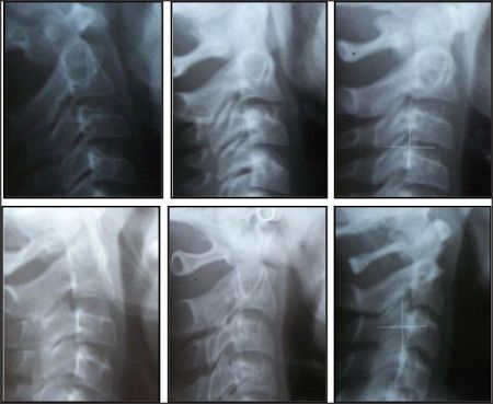 | Figure 1 - Lateral head films showing various stages of cervical vertebrae maturation- CS 1 to CS 6
 |
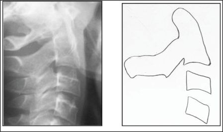 | Figure 2 - Radiograph showing patient in CS 3 with corresponding tracing.
|
Left hand wrist radiographs were evaluated by direct reading(Figure -3) of radiographs using Bjork Grave and Brown method[13] to evaluate the skeletal maturational status.
Lateral cephalograms were traced (Figure -2) for cervical vertebrae 2, 3 and 4, on acetate tracing sheet and evaluated using modified CVM method as given by Baccetti et al[14](Figure -1) to assess the level of cervical vertebrae maturation.
Statistical Analysis was performed using the Statistical Package for Social Sciences (SPSS) version 13.
Correlation among various methods was determined using x2 test and significance was established with P value set at 0.05.
Correlation values for various variables were calculated by using Spearman Correlation Coefficient (p).
Percentage distributions for different variables were evaluated
Results
Tables Igive the correlation values of CVM methods and hand wrist method. As the descriptive data of various stages was similar for males and females, the
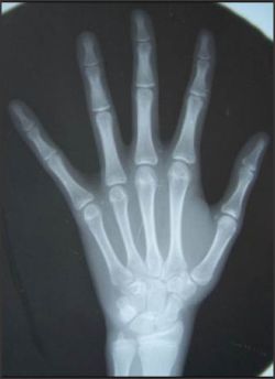 | Figure 3 - Hand wrist radiograph showing stage 8 of skeletal maturity.
|
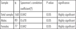 | Table I-Correlation coefficient between hand wrist maturation and cervical vertebrae stages
|
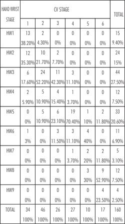 | Table II – Percentage distribution of cervical vertebrae stages among hand wrist stages for overall sample
|
percentage distribution of cervical vertebrae stages among various hand wrist stages is combined and shown for the total sample in Table II.
The spearman correlation coefficients were highly significant for comparisons among hand wrist stages and cervical vertebrae stages for both females and males.
Females showed a higher correlation with of cervical vertebrae maturation with hand wrist stages (0.842) than males (0.678). The correlation between hand wrist stages and cervical vertebrae maturation for the total sample was 0.821.
The highest percentage distribution was shown by CS 4 in HW 5 (70.4%) as shown by Table II.
Discussion
Various biologic indicators have been used to assess an individual's skeletal maturity. Hand wrist evaluation has proved to be a reliable indicator of somatic maturity. However, the routine use of hand wrist radiographs has lately been questioned due to radiation safety point of view. Thus, the use of maturity indicators observed routinely on pretreatment orthodontic records has become imperative. One such indicator available at disposal of an orthodontist is cervical vertebrae maturation status as seen on a lateral cephalogram. The aim of this study was to correlate hand wrist method and cervical vertebrae evaluation as skeletal maturity indicators and prove the efficacy of the cervical maturation status as a suitable alternative to hand wrist radiographs and thereby, protecting our patients from unjustified radiation exposure.
Hand wrist and cervical vertebrae evaluation
The results of this study (Table I) showed a highly significant correlation between hand wrist and cervical vertebrae maturation with the spearman correlation coefficient (p), of 0.84 for females (p<0.001) and 0.67 for males (p<0.001), and overall correlation coefficient of 0.82 (p<0.001) .This was in accordance with the results of Roman et al [15] who showed higher correlation coefficients for females. Thus, the use of cervical vertebrae as an indicator of skeletal maturity was found to be more reliable in females as compared to males.
The high level of correlation among hand wrist and cervical vertebrae maturation shown in this study was in accordance with Mir et al[10], Kucukkeles et al [16] and Garcia et al [17]. The value of correlation coefficients differed among these and the present study, probably due to the use of different methods of evaluation of hand wrist and cervical vertebrae. Gandiniet al[12] also showed the concordance of 83.3% in Bjork intervals and the improved cervical vertebrae maturation method.
Descriptive analysis (Table II) of the present study showed results to be in accordance with the results of Gandini et al [12].
CS 1 consisted of hand wrist stages 1, 2 and 3 (38.2%, 35.3% and 17.6% resp.)
For CS 2 more than 50% individuals were in hand wrist stage 3 followed by hand wrist stage 2 (21.7%).
Thus, 83.7% of individuals (67 out of 80) belonging either to CS 1 or 2 were in hand wrist stages 1, 2 or 3, as shown by Gandini et al[12]where CVMS 1 (CS1 and CS2 combined of present study) consisted of Bjork's hand wrist stages 1, 2 and 3.
CS 3 of the present study showed a varied distribution among hand wrist stages 3, 4, 5 and 6 with majority in stage 3 (42.3%), which is in contrast to the results of Gandini et al [12] where the equivalent cervical stage was related to hand wrist stage 4.
CS 4 had 70.4% individuals belonging to hand wrist stage 5 and relatively lesser percentage (11.1%) in stage 6.
CS 5 showed distribution among hand wrist stage 6 (40%), 7 (20%) and 8 (30%).
CS 6 represented a combination of hand wrist stages 8 (52.9%) and 9 (23.5%).
The highly significant correlation (p<0.001) of cervical vertebrae maturation stages to the hand wrist maturity stages makes it a reliable indicator of an individual's skeletal maturity. The specific distribution of hand wrist stages among the various cervical stages makes the cervical vertebrae maturation method a clinically reliable method of determining ones skeletal maturity, to assess the status of an individual's pubertal growth spurt in relation to his/her own growth cycle, with the use of a single routine pretreatment lateral cephalogram.
Conclusion
Modified Cervical Vertebrae Maturation stages can be used as a reliable indicator of an individual's skeletal maturity as they showed very high correlation with hand wrist stages and the specific distribution of hand wrist stages among the various cervical vertebrae stages provide the means to assess the status of an individual's pubertal growth spurt in relation to his/her own growth cycle, with the use of a single routine pretreatment lateral cephalogram. Thereby providing us with the means to protect our patients from unwarranted radiation exposure by eliminating the use of hand wrist radiographs.
Acknowledgement
I would like to acknowledge Dr. Pramod Philip and Dr.NavneetArora for the support and guidance provided by them in carrying out this study.
References
1. Nanda R. The rates of growth of several facial components measured from serial cephalometric roentgenograms. Am J Orthod 1955; 41:658-673
2. Hunter CJ. The correlation of facial growth with body height and skeletal maturation at adolescence. Angle Orthod. 1966; 36:44-54.
3. Greulich W, Pyle S. Radiographic Atlas of Skeletal Development of Hand and Wrist. Stanford, California: Stanford University Press; 1959
4. Bjork A, Helm S. Prediction of the age of maximum pubertal growth in body height. Angle Orthod. 1967; 37:134-143.
5. Tofani M. Mandibular growth at puberty. Am J Orthod. 1972; 62: 176-194.
6. Lewis AB, Garn SM. The relationship between tooth formation and other maturational factors. Angle Orthod. 1960;30:70-77
7. Hagg U, Taranger J. Maturation indicators and the pubertal growth spurt. Am J Orthod. 1982; 82:299-309.
8. Lamparski D. Skeletal Age Assessment Utilizing Cervical Vertebrae. [Master's thesis]. Pittsburgh, Pa: University of Pittsburgh; 1972. As cited by O'Reilly MT, Yanniello GJ. Angle Orthod. 1988; 58:179-184.
9. O'Reilly MT, Yanniello GJ. Mandibular growth changes and maturation of cervical vertebrae-a longitudinal cephalometric study. Angle Orthod. 1988; 58:179-184.
10. Rajagopal R, Kansal S. A comparison of modified MP3 stages and cervical vertebrae as growth indicators. J ClinOrthod 2002;36:398-406
11. Carlos Flores-Mir, Corr A. Burgess, Mitchell Champney, Robert J. Jensen, Micheal R. Pitcher, Paul W. Major. Correlation of Skeletal Maturation Stages Determined byCervical Vertebrae and Hand-wrist Evaluations.Angle Orthod 2006; 76:1-5.
12. Gandini P, Mancini M, Federico. A comparison of hand wrist bone and cervical vertebral analyses in measuring skeletal maturation. Angle Orthod 2006; 76:984-989
13. Grave KC, Brown T. Skeletal ossification and the adolescent growth spurt. Am J Orthod. 1976; 69:611-619
14. Baccetti T, Franchi L, and James A. McNamara. The Cervical Vertebral Maturation (CVM) Method for the Assessment of OptimalTreatment Timing in Dentofacial Orthopedics. SeminOrthod 2005;11:119-129.
15. San Roman P, Palma JC, Oteo MD, Nevado E. Skeletal maturation determined by cervical vertebrae development. Eur J Orthod. 2002; 24:303-311
16. Kucukkeles N, Acar A, Arun T. comparisons between cervical vertebrae and hand wrist maturation for the assessment of skeletal maturity. J ClinPediatr Dent 1999; 24:47-52. As cited by Grave K, Townsend G AustOrthod J. 2003; 19:33-45.
17. Garcia-Fernandez P, Torre H, Flores L, Rea J. The cervical vertebrae as maturational indicator. J ClinOrthod1998; 32:221-225. |