Introduction
According to glossary of prosthodontic shemisection is defined as, "the surgical separation of a multi-rooted tooth, especially a mandibular molar, through the furcation, in such a way that a root and associated portion of the crown may be removed."[1]
Buhler stated that hemisection should be considered before every molar extraction because this procedure can provide a good absolute and biologic cost saving alternative with long term success.[2]
Various other resection procedures are: root amputation, radisection and bisection. Root amputation refers to removal of one or more roots of multirooted tooth while other roots are retained. Radisection is a newer terminology for removal of roots of maxillary molars. Bisection / bicuspidization is the separation of mesial and distal roots of mandibular molars along with its crown portion, where both segments are then retained individually.[3]
Hemisection of either a maxillary or mandibular molar is often a means of retainingteeth needed for restorative abutments or occlusal support. Periodontal, prosthodontic and endodontic assessment for appropriate selection of cases is important for the predictable outcome.[4]
This case report describes hemisection procedure which was chosen to retain the endodontically treated mesial root of mandibular left first molar and extraction of periodontally involved distal root followed by placement of single crown over mesial root to restore the space.
Indications and Contratindications For Hemisection
According to Weine, [3] indications for root amputation includes :
Severe bone loss limited to one root or involvement of a class III furcation that could produce a stable root after hemisection.
Inappropriate performance of oral hygiene by the patient in the area.
Extensive exposure of the roots because of dehiscence is another indication for excision of one root.
Failure of an abutment within a fixed prosthesis provided a portion of the tooth can be retained to act as the abutment for the prosthesis.
Untreatable endodontic failure due to perforations and broken instruments.
Vertical root fracture confined to a single root of a multirooted tooth or any severe destructive process that is confined to a single root, including caries, external root resorption and trauma.
According to Ingle, [5]contraindications for root amputations includes:
Lack of necessary osseous support for the remainingroot or roots.
Fused roots or roots in unfavorable proximity toeach other.
Remaining root or roots endodontically inoperable.
Lack of patient motivation to properly performhome-care procedures.
Case Report
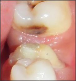
A 28 years old female patient reported with thecomplaint of pain and mobility of left mandibularfirst molar. Patient did not give any significant medical and previous dental history. Extra oral examination revealed no abnormality. On intraoral examination, it was found that patient had fair oral hygiene with grossly decayed distal surface of 36 (Fig 1). On probing lower left mandibular first molar, a periodontal pocket of 8-10mm was found on distal root surfaces along with
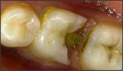 | Fig 1
 |
grade III furcation involvement. The tooth wassensitive to percussion and revealed grade Imobility. On radiographic examination, severevertical bone loss was evident surrounding thedistal root and involving the furcation area. Thebony support of mesial root was completely intact. Periodontal prognosis with 36 was good and vitality test was positive. Thus, it was diagnosed as chronicgeneralized gingivitis and localized periodontitis associated with lower left mandibular first molar. Treatment options included extraction of 36 followed by placement of implant, fixed partial denture or removable partial denture. Patient did not wish to have the tooth removed, so it was decided that the distalroot of 36 should be hemisected after completion ofendodontic therapy of the tooth followed by prosthetic replacement.
According to Rosensteil et al, [6]the most common types of restorations for teethwith resected roots involve:
1. The remaining root restored as an individual tooth.
2. The tooth used as an abutment for a fixed orremovable partial denture
3. Premolarization-individual roots of a molarrestored with premolar morphology
4. Minimum treatment-amalgam placed in theroot(s) and the occlusion adjusted
It was decided to choose the option 1.
The Endodontic Phase
Endodontic phase involved intentional root canal treatment of 36 in a conventional manner (Fig 2).The working length was determined and the canals were biomechanically prepared using stepback technique. The canals were obturated with lateral condensation method and the chamber was filled with composite. After 15 days ofobturation, hemisection was carried out.
The Periodontic Phase
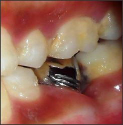
After appropriate local anaesthesia, a crevicular incision wasmade from first premolar to second molar region. A fullthicknessmucoperiosteal flap was elevated. After reflection of flap, bonydefect was evident and curettage and debridement weredone. A long shank tapered fissure carbide bur was usedto make vertical cut facio-lingually towards the bifurcationarea and distal root was extracted (Fig 3). Care was taken notto
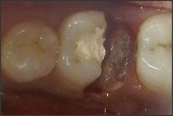 | Fig 2
|
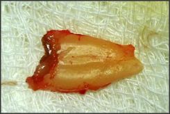 | Fig 3
|
 | Fig 4
|
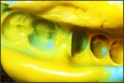
traumatize bone and adjacent tooth while removing thedistal root. Debridement and irrigation of the socket alongwith thorough root planning of mesial root was performed. Odontoplasty was performed to remove the developmental ridges, and distal aspect of mesial root was contoured insuch a way so as to facilitate oral hygiene measures. The extraction site was irrigated and debrided and the flap was then repositioned and sutured. The surgical site was then allowedto heal with no occlusal stress on distal root for 4 weeks.Patient was recalled after 3 months. Then, the restoration of hemisected tooth was planned with using the remaining tooth portion to be restored as as an individual tooth as the space for the pontic of FPD was very less (Fig 4).
The Prosthodontic Phase
Diagnostic impressions were made with irreversible hydrocolloid impression material and diagnostic casts wereobtained. Face bow record was made and transferred to a semi-adjustable articulator and maxillary cast was mounted. Mandibular diagnostic cast was mounted using interocclusal record.
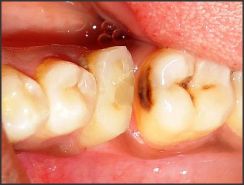
Tooth preparation was done in relation to 36 to receive a metal restoration to look alike the molar. (Fig 5)
Final impression was made using putty-reline technique and master cast was obtained.(Fig 6) Mandibular master cast was mounted using interocclusal record.
Wax pattern was fabricated in a manner so that it appear as mandibular first molar occlusally but contacting the opposing dentition only on the mesial half, distally the wax pattern was extended till the mesial contact point of 37 to prevent mesial tilting; the distal embrasure was kept wide to allow adequate oral hygiene manually; the wax pattern was thensprued, and invested.
Casting procedure was carried out using standard techniques.
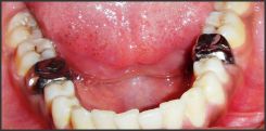
Metal coping was tried in the patient's mouth.Occlusion was checked to remove any contact on disto-occlusal surface to prevent lateral forces on mesial root. Final finishing and polishing were carried out. (Fig 7).
Final prosthesiswas cemented using glass ionomer cement (Fig 8). Post cementation instructions regarding periodontalmaintenancewere given.
 | Fig 5
|
 | Fig 6
|
 | Fig 7
|
 | Fig 8
|
Recall was done periodically to assure thehealing and success of the restoration.
Discussion
The prognosis of root resected molars may not be as poor as previously believed. Multirooted, periodontally involved molars can be maintained for long periods of time with hemisection. Thelarge variation in success and failure reported by different authors is a reflection that roots resection and hemisection is a technique sensitive procedure. One must be careful throughout the processes of case selection, and endodontic, periodontal, restorative and maintenance therapies. Critical analysis before reconstruction and regular reevaluation during maintenance period are crucial.[7]
The treatment options to replace severely damaged and possibly unrestorable teeth include removable partialdenture, fixed partial denture, and dental implant. A guiding principle should be to try and maintain what is present. The use of hemisection to retain a compromised tooth offers a prognosis comparable to any other tooth with endodontic treatment.[8]
When choosing to perform a hemisection procedure, consideration should be given to the morphology, clinical length and shape of the roots of a multirooted tooth. It is important to take into account the divergence of the roots while making a case selection. Affected teeth with roots spread apart facilitate the clinician's ability to carry out root resection. Teeth with closely approximated or fused roots are not good choices to receive hemisection therapy.[4]
In the present case the above mentioned indication for case selection in performing hemisection was optimum as the roots were not closely approximated or fused.
Hemisection has been used successfully to retain teeth with furcation involvement. However, there are few disadvantages associated with it. As with any surgical procedure, it can cause pain and anxiety. Root surfaces that are reshaped by grinding in the furcation or at the site of hemisection are more susceptible to caries. Often a favorable result may be negated by decay after treatment. Failure of endodontictherapy due to any reason will cause failure of the procedure.[9]
When the toothlose part of its root support, it will require a restoration to permit it to function independently or serve as an abutment for fixed partial denture or splint. Thus, restoration is required for function and stabilization of occlusion. Points to consider while fabricating the prosthesis: restoration can contribute to periodontal destruction, if margins are defective or if nonocclusal surfaces do not have physiologic form. An improperly shaped occlusal contact area converts acceptable forces into destructive forces leading to ultimate failure of hemisection. Occlusal table is reduced in size in order to decrease the forces on the retained hemisected root. Cuspal inclines are made less steep to reduce laterally directed forces and eliminate the nonworking contacts. Retained root is restored as premolar which helped to reduce the masticatory load. Stein noted that "esthetic permitting, the sanitary pontic is the best design for posterior region". [10]
Thus, this article presents a technique for the dentist to restore the hemisected mandibular molar with single crown in cases where there is very less space forpontic by keeping the occlusal contact only on the mesial half of the 36 to prevent the torquing forces on the root.
Conclusion
The keys to long term success include thorough diagnosis, selection of patients with good oral hygiene, careful surgical and restorative management. Hemisection may be a suitable alternative to extraction and implant therapy and should be discussed with patients during consideration of treatment options.with recent refinements inendodontics, periodontics and restorative dentistry, hemisection has received acceptance as a conservative and dependable dental treatment and teeth so treated have endured the demands of function.
References:
1. The Glossary OfProsthodontic Terms. J Prosthet Dent 2005; 94:41.
2. Buhler H. Survival Rates Of Hemisected Teeth: An Attempt To Compare Them With Survival Rates Of Alloplastic Implants. IntJPeriodontics Restorative Dent. 1994; 14: 536-543.
3. Weine FS. Endodontic Therapy.5th St Louis: Mosby; 2002.
4. Kurtzman GM, Silverstein LH, Shatz PC. HemisectionasAn Alternative Treatment For Vertically Fractured Mandibular Molars. CompendContinEduc Dent 2006; 27(2):126-9.
5. Ingle JI, Bakland LK. Endodontics. 6th Ed. London: Mosby; 2006.
6. Rosenstiel SF, Land MF, Fujimoto J. Contemporary Fixed Prosthodontics. 4th Ed. Edinburgh: Elsevier Mosby; 2006.
7. Baston CH, Ammons WF, Persson R. Long Term Evaluation of Root Resected Molars: A Retrospective Study. Int J PeriodontRestor Dent 1996; 16(3):207.
8. A. T. Nowakowski, A. Serebnitski, and I. J. Pesun, "Hemisectionas A Treatment Option: A Case Report," Oralhealth2010;100(3): 83-89.
9. G. Parmarand P. Vashi, "Hemisection: A Case Report and Review," Endodontology 2003; 15: 26-29.
10. M. S. Schmitt and H. F. Brown."The Hemisected Mandibular Molar. A Strategic Abutment," J ProsthetDent1987; 58(2): 140-144. |