Introduction:
Anodontia is the congenital absence of teeth and can occur in some or all teeth (partial anodontia or hypodontia), involve two dentitions or only teeth of the permanent dentition (Dorland's 1998). Many denominations are attributed to this anomaly: partial anodontia, hypodontia, oligodontia, the congenital absence, anodontia, bilateral aplasia. Anodontia being the term used in controlled vocabulary Medical Subject Headings (MeSH) from MEDLINE which was developed by the USA National Library of Medicine. The Anodontia of at least one permanent tooth is the most common dental anomaly and may contribute to masticatory dysfunction, speech impairment, aesthetic problems, and malocclusion [1]. Absence of lateral incisors represents a major stereotype. Individuals with this condition are perceived as socially most aggressive compared with people without anodontia (Shaw 1981) [2].Partial anodontia, known as hypodontia or oligodontia, is the congenital absence of one or more teeth. Congenital absence of all wisdom teeth, or third molars, is relatively common
Case Report:
Case 1
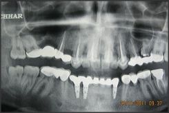
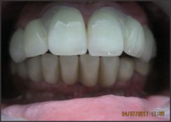
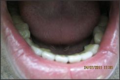
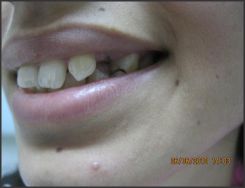
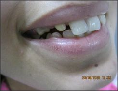
A 24 year old male patient came with his sister who is 26 years old with a complaint of poor esthetics. During the history taking the male patient revealed that he had got done root canal treatment and PFM crowns on the Maxillary Central Incisors and canines on both the sides few years back from elsewhere and that the lateral incisors never erupted so that area was restored with the bridges (Fig. 1 & 2). On examination, there was gingivitis in relation to faulty bridges, midline diastema which could not be closed by the bridges and also the crowns were bulky and esthetically not acceptable. On examination it was observed that there was an edentulous area in the mandibular anterior region from canine to canine. Patient didn't give any history of extraction in that region and also mentioned that the teeth never erupted in that region. All the findings were confirmed with the help of OPG (Fig. 3) .The study models revealed the absence of maxillary second premolars and retained deciduous molars in both the maxillary quadrants ,in the mandibular arch the anteriors from canine to canine and also both the second premolars were
 | Fig 1 - Preoperative Photo - Anterior View
 |
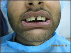 | Fig 2 - Clinical Picture Showing Missing Teeth
|
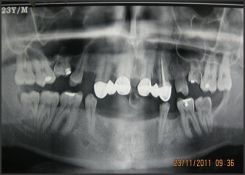 | Fig 3 - OPG Showing The Missing Teeth
|
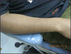 | Fig 4 - Photo Showing Sparse Hair Growth On The Forearm
|
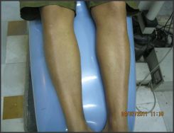 | Fig 5 - Photo Showing Sparse Hair On The Legs
|
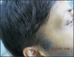 | Fig 6 - Photo Showing Receded Hairline
|
 | Fig 7 - OPG Showing Complete Rehabilitation
|
 | Fig 8 - Clinical Picture Showing Post Operative Rehabilitation
|
 | Fig 9 - Photo Showing The Lower Posteriors After Rehabilitation
|
missing. On physical examination, scanty hair growth was observed on the limbs (Fig. 4 and 5), although the scalp had enough hair but the hairline appeared to be receded and sidelocks not well defined (Fig. 6).The other commonly associated symptoms with oligodontia related to smell skin (ectodermal dysplasias), sweat glands and nerves appeared to be absent which are generally part of a syndrome associated with oligodontia.
Treatment:
To begin with 3 implants were placed in the lower anterior region, which was edentulous, after careful examination of the ridge area. Then we started with other restorations leaving the implants for at least 12 weeks to allow osseointegration (Fig. 7). In the next step we started with the replacement of maxillary anterior crowns. Metal free crowns were planned for the maxillary anterior region for better esthetics (Fig. 8). After cementation of the crowns we started with the extraction of the retained primary molar in both the upper arches. During the extraction of right upper primary molar we realised that they were ankylosed, so somehow we extracted the right one but decided to avoid extraction of the left one. After doing Root Canal Treatment for all the permanent premolars and molars in the maxillary arch we prepared the teeth for bridges.PFM crowns were planned to cut down the cost of treatment. After cementation of the maxillary bridges we prepared the existing teeth in the mandibular arch for bridges. Later after the wait period of 3 months the mandibular bridge was cemented over the implants (Fig. 9).
Case 2
On taking the history of the female patient who is the blood sister of the above mentioned male patient, she complaint of the absence of maxillary lateral incisors and canines present in place of lateral incisors. The canine space was edentulous in both the maxillary quadrants which were aesthetically not acceptable (Fig. 1 and 2). Patient gave the history of extraction of right lower first molar 5 years ago .On general examination no other symptom was observed relating to the syndrome associated with hypodontia.
Treatment planning was done for the lady keeping in mind the age factor, the esthetics and the longevity of the treatment result. A bridge on both the sides of the maxillary arch was planned after doing the root canal treatment of the
 | Fig 1 - Photo Showing Canine In Place Of Missing Lateral Incisor
|
 | Fig 2 - Prepared Canine And Premolar Before Prosthesis
|
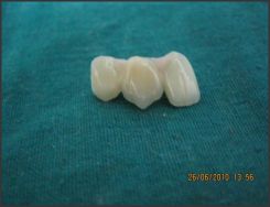 | Fig 3 - All Ceramic Prosthesis Before Luting
|
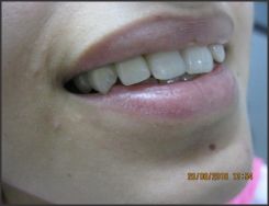 | Fig 4 - Prosthesis After Luting
|
 | Fig 5 - OPG Showing The Bridges Bilaterally
|
canines and the first premolars. The canine was reduced to the size of reduced lateral incisor to receive a lateral incisor crown and the pontic was given a shape of a canine and a premolar over a premolar. A metal free zirconia bridge was cemented after doing the root canal treatment of both the teeth on both the sides of the maxillary arch (Fig. 3, 4 & 5kll). Patient didn't want the replacement of right mandibular molar as of now. This lady didn't apparently show any other signs and symptoms on the body anywhere related to hypodontia.
Discussion:
Anodontia is the congenital absence of all teeth. Edentulous means the clinical absence of teeth. Anodontia means the failure of teeth to form at all. In true (or complete) anodontia, all teeth fail to develop.
-True anodontia is an extremely rare occurrence. When this does occur, it is usually part of a more generalized disorder ectodermal dysplasia, an inherited defect of all ectodermally derived structures.
-Partial anodontia is the failure of one or more teeth to develop. It is much more common than complete anodontia. Third molars, lower second premolars, and upper lateral incisors (in that order) are the most common congenitally absent teeth. (Note this very well: congenital absence of a deciduous tooth is not common. When it does occur, it is most often the upper lateral incisor that is absent. When a deciduous tooth is absent, its permanent successor is usually missing also).
-Supernumerary teeth are 'extra' teeth. Most (90%) occur in the maxilla. Their presence in the deciduous dentition is quite rare. Two terms occasionally used to describe this condition of having extra teeth are polydontia or hyperdontia. The most common of all supernumerary teeth is the mesiodens which is a supernumerary that forms in the midline between and lingual to the roots of the maxillary central incisors. Most supernumerary teeth do not erupt; they often are unknown until detected on X-ray films. Fourth molars, themselves a rare occurrence are infrequently called 'paramolars' or 'distomolars.' Supernumerary teeth occur less often than do missing teeth. Congenitally missing teeth (hypodontia and oligodontia) are not rare. . With the discovery of the Pax 9 gene, some light has been shed on the molecular genetics of congenitally missing teeth. Pax 9 maps to chromosome #14, it encodes a transcription factor that functions in the development of derivatives of the pharyngeal pouches. Mice with Pax 9 mutations have "craniofacial and limb anomalies and teeth fail to form beyond the bud stage". A family with a framshift mutation in Pax 9 had normal deciduous teeth but lacked permanent molars in both the maxilla and mandible. It is transmitted as a dominant trait. [3], [4], The best known of the missing teeth syndromes is X-linked hypohidrotic ectodermal dysplasia. Ectodermal derivatives such as hair, sweat glands, nails and teeth are involved. Head hair, eye lashes and brows are sparse, nails are dystrophic and there is marked oligodontia, rarely total anodontia. Diminished sweat glands lead to the inability to regulate body temperature, a major disability in warm months in a hot climate. According to your text, there are 150 variants of this syndrome [5]. X-linked disease is milder in females because they enjoy partial protection thanks to lyonization. If both parents have "peg laterals", the homozygous child will have total anodontia of succedaneous teeth. [6]
Partial anodontia. Congenitally missing teeth are more common among individuals with Down syndrome (50%) when compared to the general population (2%), though the distribution of missing teeth is similar in both populations.[7], [8] Genetic modes of transmission are responsible for this condition. A relationship between partial anodontia and other ectodermal defects (mucous membrane, hair, skin) has been suggested.[9], [10], [11] Further research indicated that this "Trisomic insult" will greatly increase the susceptibility of the host to partial anodontia, while not affecting specific tooth buds. [7], [8] The most frequently missing teeth in descending order are third molars, second bicuspids, lateral incisors, and mandibular incisors. The only teeth not missing are first molars. [7], [8]. Sometimes the primary tooth will not be resorbed or will be resorbed so slowly that it can be retained well into adulthood [12]. Treatment decisions should be made after reviewing the radiographs with concern for space maintenance. In the general population, the incidence of supernumerary primary teeth is approximately 0.3% while in the Down syndrome population the incidence is increased but the frequency is less than congenitally absent teeth. Crowding is not uncommon and seen more in the maxillary arch than the mandibular arch[12].
Conclusion:
Ectodermal dysplasia along with partial anodontia may not occur eveytime in everyone .It is more dependent on the genetic structural changes. Like in the cases discussed above where the siblings were affected from partial anodontia but only the male was affected with ectodermal dysplasia .
References:
1. Shapiro SD, Farrington FH. A potpourri of syndromes with anomalies of dentition. Birth Defects Orig Artic Ser. 1983;19(1):129-40.
2. Shaw, W. The influence of children's dentofacial appearance on their social attractiveness as judged by peers and lay adults. American Journal of Orthodontic.s 1981; 79(4): 399--415.
3. David W. Stockton , Parimal Das , Monica Goldenberg , Rena N. D'Souza & Pragna I. Patel. Mutation of PAX9 is associated with oligodontia. Nature Genetics 2000; 24:18-19.
4. Peters H, Neubüser A, Balling R. Pax genes and organogenesis: Pax9 meets tooth development. European Journal of Oral Sciences 1998; 106: 38-43.
5. Neville's Oral & Maxillofacial Pathology 2nd edition.
6. Carl J. Witkop Jr. and James F. Reynolds. Agenesis of succedaneous teeth: An expression of the homozygous state of the gene for the pegged or missing maxillary lateral incisor trait. American Journal of Medical Genetics 1987; : 431-436.
7. Desai S S. Down Syndrome: A Review of the Literature. Oral Surgery, Oral Medicine, Oral Pathology, Oral Radiology, and Endodontics; 1997; 84 (3); 279-85.
8. Scully C. Down syndrome: aspects of dental Care. J. of Dent 1976; 4:167-74.
9. Wilson MD. Special considerations for patients with Down syndrome. ODA J 1994; l84 (3):24-25.
10. Hay RJ, Wells RS. The syndrome of ankyloblepharon, ectodermal defects and cleft lip and palate: an autosomal dominant condition. American Journal of Medical Genetics 1976; 94 (3), 277-289.
11. Torfs CP, Christianson RE. Anomalies in Down syndrome individuals in a large population-based registry. American Journal of Medical Genetics 1998; 77(5): 431-438.
12. Limbrock G., Fischer-Brandies H., Avalle C. Castillo-Morales' orofacial therapy: treatment of 67 children with Down syndrome. Dev Med Child Neurol. 1991: 33:296-303.
|