Introduction
The disfigurement associated with the loss of an eye can cause significant physical and emotional problems (Lubkin & Solan, 1990). The rehabilitation of a patient who has suffered the psychological trauma of an ocular loss requires a prosthesis that will provide the optimum cosmetic and functional result. Refinement in the details of custom made ocular construction has produced a superior restoration delivered more readily[1].
Depending on the severity of the situation, the management may include 3 approaches which need the combined efforts of the ophthalmologist, the surgeon, and the maxillofacial prosthodontist to restore the patient's quality of life[2].
The surgical procedures in the removal of an eye are classified into three categories, namely evisceration, enucleation and excenteration. Evisceration is the removal of contents of the eye ball leaving behind the sclera. After evisceration, a plastic conformer and corticosteroid antibiotic ointment is placed in the socket. The plastic conformer is left in place for 4-6 weeks to reduce edema and maintain the socket contours for a prosthetic eye. When surgical site is well healed and dimensionally stable, fabrication of an ocular prosthesis may be undertaken. Early management of anophthalmic socket prevents loss of volume in the anterior orbital area and facial asymmetry [3]. Various methods of rehabilitating an anopthalmic socket include, a) stock eye prosthesis (Prefabricated), b) custom made ocular prosthesis [4].
In this case report an easy and economical method of fabricating an ocular and scleral prosthesis is described.
Case Report
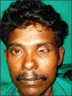
A 28 year old male patient reported to the Department of Ophthalmology, K.V.G. Medical College and hospital with a chief complaint of lack of vision and pain in the left eye. Patient gave a history of trauma from a hay stick while working in field, and had not taken any medical treatment. Visual perception for light was nil but had intact eye movements. The case was diagnosed as corneal abscess with endopthalamits (Fig 1).
Treating this case needed surgical removal of the corneal contents and rehabilitation with prosthesis. Ophthalmologist and Prosthodontist, with an Interdisciplinary approach made the following treatment plan. Rehabilitation was planned in two
 | Fig VII Post operative
 |
 | Fig VII Post operative
|
 | Fig VII Post operative
|
 | Fig VII Post operative
|
phases. In first phase, evisceration of the intraocular contents and placement of heat cured Poly (methyl methacrylate) [PMMA], ball implant prosthesis during the time of surgery . In second phase after 4-6 weeks of surgery fabrication of custom made scleral prosthesis .
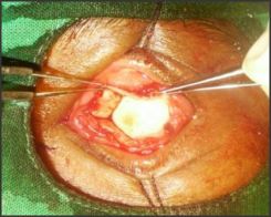
A 18 mm heat cured poly methylemethacry late ball was implanted inside the hollow cavity (Fig 2). Tenon's capsule was sutured with 6-0 vicryl
 | Fig VII Post operative
|
suture and the conjunctiva with nylon suture. The operated eye was allowed to heal for a period of 6 weeks.
Primary impression was made using irreversible hydrocolloid (Zelgan 2002; Dentsply DeTrey GmbH,Konstanz, Germany). The patient's eye socket was coated with thin layer of Vaseline and impression material was injected into the depth of upper and lower eye lids, while making impression patient was asked to eye movement so that functional impression of the defect could be obtained. The impression was then retrieved and poured to obtain a primary sectional cast and special tray with clear acrylic was fabricated.
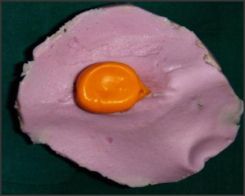
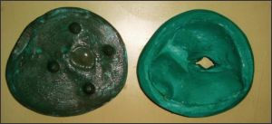
Final impression was made using light body polyvinyl siloxane addition silicon (Reprosil;Dentsply India), it was injected using 5ml syringe, at the end of syringe perforated special tray was attached(Fig 3). Irreversible hydrocolloid impression material was placed over the light body material; over it type II gypsum material was poured to reinforce it. Impression was poured by multiple pour technique and sectional cast was obtained (Fig 4).
Then the two halves of casts were separated and the final impression retrieved so as to keep the mold space ready for wax pattern fabrication for which molten wax was poured into the secondary cast through the sprue channel. When the wax had set the cast was separated and the wax pattern was retrieved and carved. This wax pattern was tried in patients socket and lid contours, eye movement, and retention were evaluated, once satisfied, it was invested with tooth color heat cure acrylic. Once acrylic sclera prosthesis finished and polished, it's reevaluated in patient's eye for fit, contour, and retention.
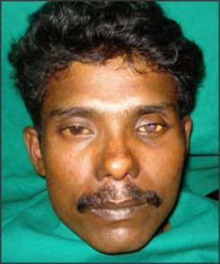
Iris was fabricated using paper iris disk technique. Fine red threads of the veined heat cure acrylic were used to mimic the blood vessels of the patient's natural eye. Oil color was also used over the scleral blank to mimic the adjacent eye. The scleral blank was flasked and clear acrylic resin was added and trial packed. Once after verifying that the iris is not moved, the flask was closed and then processed. Then the prosthesis was finished, polished and delivered to the patient. The patient was educated to insert and remove the prosthesis and post insertion instructions were given. The final outcome of the prosthesis was ascertained from the satisfied look on the face of the patient and patient was called for routine follow up (Fig 5).
Discussion
The rehabilitation of a patient who has suffered the psychological trauma of an ocular loss requires a prosthesis that will provide the optimum cosmetic and functional results. Ocular prosthesis has a long history of successful use, and variation of the techniques and materials used have been introduced throughout the years [5].
An correctly placed ocular prosthesis should maintain its orientation when the patient is looking straight ahead. A correctly placed prosthesis should restore a degree of movement, normal opening of eye, support the eyelids, and be adequately retained with esthetic appearance [6].
In this paper disk technique, digital photography is used to replicate the iris of the patient, replacing the conventional oil paint and monopoly iris painting technique. Advantage such as reduced treatment time and increased simplicity make this method an alternative for fabricating ocular prosthesis.
Laney and Gardner have been advocated use of stock ocular prosthesis of an appropriate size and color , adapted by selective grinding and addition of acrylic resin can give excellent results for most of patients, provided the operator has an adequate selection of prefabricated scleral prosthesis and experience[7]. However, because of the extreme individual anatomical variation and diverse nature of ocular injuries, patients would benefit more from custom made ocular prosthesis that are modified to their individual needs. This procedure may be more time consuming and entail a "trial and error" approach, but the esthetic and functional results justify the extra efforts.
Conclusion
Most patients experience significant stress, due primarily to adjusting the functional disability caused by the eye loss, and to social reaction to the facial impairment. Replacement of lost eye as soon as possible after healing from surgery is necessary to promote physical and psychological healing for the patient and to improve the social acceptance. A multidisciplinary approach and team work are essential in providing accurate and effective rehabilitation of such patients.
The use of custom made ocular prosthesis has been a boon to the patient who cannot afford for the implant placement. An ocular prosthesis does not provide vision; but this would be a visual prosthetic, which has definitely restored patient's self-esteem and allowed him to confidently face the world rather than hiding behind dark glasses.
References
1. Lubkin V, Sloan S: Enucleation and psychic trauma. Advances in Ophthalmic Plastic and Reconstructive Surgery, 1990; 8:259-262.
2. Thomas D Taylor. Clinical maxillofacial Prosthesis. Quintessence Publishing Co;2000;233-76.
3. Baylis H, Shorr N, McCord C. Evisceration, enucleation, and exenteration. Oculoplastic Surgery 2nd ed New York: Raven 1987: 425-49.
4. Su GW, Yen MT. Current trends in managing the anophthalmic socket after primary enucleation and evisceration. Ophthalmic Plastic & Reconstructive Surgery 2004; 20(4):274.
5. Taicher S, Steinberg HM, Tubiana I, et al. Modified stock ocular prosthesis. J. Prosth. Dent. 1985;54:95-8.
6. L.M. Skyes, BSc, BDS, MD0ent. Custom made ocular prosthesis: A clinical report. J. Prosth. Dent. 1996;75:1-3.
7. Laney WR, Gardner AF. Maxillofacial prosthesis. Littleton: P S G Publising, 1979:255-89. |