Introduction
Age estimation of an individual in forensic science is very helpful for personnel identification and to establish mortality patterns in past population. Study of age related changes in dental tissues are often used to estimate the age of an individual. It is a well-established fact that with advancing ages the size of the dental pulp cavity decrases as a result of secondary dentine deposition. The measurement of this reduction in size of the pulp chamber can be used as an indicator of age in forensic and anthropological science.
Several methods of age estimation have been studied based on morphology of human permanent dentition like tooth wear, root dentine transparency, tooth cementum annulation and deposition of secondary dentine.
Previous studies have shown that periapical radiographs may be used for estimation of age[1]. But due to interobserver difference the age calculation may be less accurate when used by different investigators. To reduce inter and intra observer bias, image analysis procedure have been suggested for measurement of morphological parameters in dental tissues[2], [3].
The purpose of the present study was to estimate the age of an individual by determining pulp/tooth ratio of maxillary canines using intraoral periapical radiographs.
Materials and Method
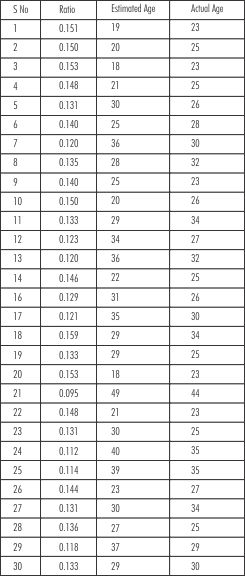
Intra oral periapical radiographs of 30 individuals whovisited our out patient department were taken randomly. All individual were of age group of 20-50 years. Intraoral periapical radiographic images of non-carious healthy maxillary canines were taken with the help of radio-visiuo graphy (RVG). The obtained digital images of canines were processed using a computer-aided drafting program (AutoCAD2000).
Inclusion Criteria
Non-carious healthy maxillary canines were included in this study. Canines were slected in this study since they are mostly present in old age; they are less likely of wear than other anterior teeth and are the single-rooted teeth with the largest pulp area, and thus easy to analyze.
Exclusion Criteria
Multirooted teeth were excluded from the present study because of the difficulty in defining the pulp in each root on a radiograph. Endodontically treated teeth were also excluded because in root filled teeth there are no functioning odontoblast and thus no further secondary dentine formation, which together with the instrumental widening of the pulp, makes the tooth unsuitable for measuring. Apical pathological processes were considered as sign of necrosis and might represent a halt in production of secondary dentine. Such teeth were excluded from the study. Totally impacted teeth remainunexposed to oral cavity and formation of secondary dentine is then a slower process than in functioning teeth. Hence impacted teeth were also excluded.
Twenty points from each tooth outline and ten points from each pulp outline were identified and used to evaluate both tooth and pulp areas. (Fig. 1) Following this pulp-tooth ratio was calculated (Fig. 2) and age was estimated using
 | Figure 1
 |
 | Fig 3
|
 | Figure 2
|
regression equation. I.e
Estimated age= 99.937-532.775(x)
Results
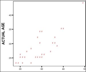
Age difference between the estimated age and actual age was +-7 years. Plotting these values on interactive graph it revealed a positive correlation between the estimated age and actual age of the subject. (Fig. 3). Followingregression equation was derived from present study.
Age= (13.196+0.528) x estimated age
Using this formula, the age difference between the estimated age and actual age was +- 2 years.
Discussion
Most studies for age estimation have used bone, which changes as an individual grows. Bone modification is visible until subject reaches adulthood, and subsequently in degeneration process[4]. A particular problem for age estimation is that premortem modification may vary from subject to subject and postmortem changes and taphonomy is influcenced by many factors (time, humidity etc). Of various parts of body used in age estimation, teeth are the least effected. Several age estimation methods involving teethstudy tooth modifications including wear[5],[6], root dentine transparency[7],[8], and tooth cementum annulation[9],[10], racemization of aspartic acid[11], [12] and apposition secondary dentine[1],[2]. Wear and apposition of secondary dentine are the currently available nondestructive methods. Tooth wear is influenced by various external factors like- masticatory function, type of food, timing and sequence of tooth eruption, tooth form, position of teeth, thickeness and hardeness of enamel and predisposition to enamel hypoplasia. All these methods require extraction, and preparation of microscopic sections of at least one tooth from each individual. These methods cannot be used in living individual and in cases where it is not acceptable to extract teeth for ethical, religious, cultural or scientific reasons. Howerver, the apposition of secondary dentine is continous, regular process, which is only modified by caries or tooth abrasion. It is a well-established fact that with advancing age the size of the dental pulp cavity decreases as a result of secondary dentine deposition. The measurement of this reduction in size of the pulp chamber can be used as indicator of age in forensic and arthopological science. The need to estimate age in skeletons of adult people is important in forensic and anthropological sciences. Although several parts of the body remains can be used for age estimation, the poor condition of the remains, often prevent their use. For this reason, the teeth are that part of the human body most frequently used for identification and age estimation when skeletal remains are in poor conditions.
Conclsion
The result of present study reveals that canines can be used as morphological variables to predict individual age in the absence of conventional sources.
References
1. S.Kvaal, K.M. Kolltvert, I.O. Thomsen,T.Solheim, Age Estimation Of Adults From Dental Radiographs, Forensic Sci. Int. 74(1995)175-185.
2. M.Lopez-Nicolas, M.Canterase, A.Luna, Age Estimation By IBAS Image Analysis Of Teeth, Forensic Sci Int.45(1990)143-150
3. L.Kollman, T.Matrinsson, The Accuracy Of Measuring Tooth Lengths From Intraoral Radiographs Using Coputerized Registration, Dentomaxillofac. Radical. 17(1998) 105-107
4. Roberto Cameriere, Luigi Ferrante Et Al. Age Estimation By Pulp/Tooth Ration In Canines By Peri-Apical X-Rays. J. Forensic Sci. January 2007, vol52, no 1
5. Brothwell D.The Relationship Of Tooth Wear To Aging. In:Iscan MY, Editor.Age Markers In Human Skeletons.Springfeild, IL:Charles C. Thomas Publishers Ltd, 1989:303-16
6. Lovejoy Co. Dental Wear In The Libben Population. Its Functional Pattern And Role In The Determination Of Adult Skeletal Age At Death. Am J Phys Anthrop 1985; 68:47-56.
7. BangG, Raman E. Determination Of Age In Humans From Root Dentine Transparency. Acta Odonto Scand 1970:28:3-35.
8. Lamendin H, Baccino E, Humbert J F, Tavernier Jc, Nossintchouk Rm. A Simple Technique For Age Estimation In Adult Corpus: The Two Criteria Dental Method. J Forensic Sci 1992;37:1373-9.
9. Wittwear-Backofen V, Gampe J, Vavpel Jw. Tooth Cementum Annulation For Age Estimation: Results From A Large Known-Age Validation Study. Am J Phys Anthropol 2004; 123:119-29
10. Jankauskas R, Barakauskas S, Bojarun R. Incremental Lines Of Dental Cementum In Biological Age Estimation. Homo 2001;52:59-71.
11. Ritz-Timme S, Cattaneo C, Collins Mj, Waite Er, Schutz Hw, Koatsch Hj, Et Al. Age Estimation: The State Of The Art In Relation To The Specific Demands Of Forensic Practise. Int J Legal Med 2000:113:129-36.
12. Ohtani S. Studies On Age Estimation Using Racemization Of Aspartic Acid In Cementum. J Forensic Sci 1995;40:805-7.
|