Introduction:
Implantation is a term used to “designate the operation of introducing either a natural or artificial tooth into an artificial socket cut into the alveolar process” (Congdon in 1915). According to Boucher, Implants are alloplastic devices which are surgically inserted into or onto jaw bone. Newton's Third Law states that there is a reaction for every action, control of which is difficult to achieve intraorally. Earlier, orthodontists used extraoral traction to reinforce intraoral anchorage. Nevertheless, patients seldom used headgears 24 hours a day -7 days a week, hence this source of anchorage was often compromised. Clinicians continue to need anchorage that displays a high resistance to displacement.
The ideal intra oral anchorage would not displace, and would require a source devoid of periodontal membrane, which tends to respond to tension and pressure allowing movement through bone. The development of small diameter titanium microimplants with specially designed heads that accept ligatures, coil springs and elastomers have helped to solve the main objections to previous implants and screws.
Biomaterials Used For Dental Implants :
Over the past several decades various biomaterials have been used for surgical implant.
Materials Mainly Used For Implant Designs Are:- [1],[2]
1) Metals And Alloys :-
a) Titanium and titanium -6 Aluminum-4 vanadium (Ti-6AI-4V)
b) Cobalt-Chromium-Molybdenum - Based Alloys
c) Iron-Chromium-Nickel-Based Alloys.
2) Ceramics:
a) Aluminum, Titanium and Zirconium Oxides
b) Aluminum oxides
c) Hydroxyapatite
3) Carbon and carbon silicon components
4) Polymers and composites:
a) Polymers.
b) Composites
5) Porous and Featured coating
a) Titanium plasma sprayed.
Osseointegration:
Osseointegration is the most important stability. The term “Osseointegration” is made of two Latin words.
Osseo meaning bone
Integration meaning the state of being combined into a complete whole.
According to Branemark “Osseointegration is the direct structural and functional connection between ordered living bone and the surface of load carrying implant”.[3]
Implants In Orthodontics
Their classification can be based on the implant morphology:
1. Implant Disc :
a. Onplants
2. Screw design
a. Orthosystem Implant System [4]
b. Spider Screw [5]
c. OMAS system [6]
d. Mini Implants [7]
e. Micro Implants
3. Plate Design
a. Skeletal Anchorage system [8]
b. Zygoma Anchorage System [9]
4. Resorbable Implants / Biodegradable Implants [10],[11]
Placement Procedure :[12]
This procedure is performed under local anesthesia.
Maxillary micro-implant sites need a 300-400 angulation to the long axis of the tooth, either buccally or lingually. Thicker mandibular cortical bone generally requires 10o-20o angulations.
When placing micro-implant in palate, the greater palatine artery and nerve must always be avoided. If micro-implant is inserted through movable soft tissue rather than attached gingival, it is preferred to use a screw without a button head, placing it completely beneath the gingival with an emerging ligature wire hook for elastic engagement. This reduces the risk of inflammation and infection. The micro-implant depends upon mechanical retention within the bone, thus requires a tight fit. A low speed contra angle with a drill 0.2-0.3 mm narrower than microscrew is used for initial entry into the bone.
It is safer to use a manual screwdriver so the clinician can feel resistance from roots and make adjustments to avoid them. Whenever resistance is encountered, withdraw the implant and redrill the bone with the pilot drill before reinserting the micro-implant. When the micro-implant fits tightly, orthodontic forces can be applied immediately.
Clinical Applications:
1. Closure of extraction space :
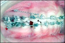
The mini screw can be placed between the roots of the first molar and the second bicuspid roots. (Figure 2)
2. Symmetric intrusion of the incisors:
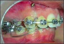
To intrude the upper incisor, the best placement of miniscrew is between the upper lateral incisors and the canines. The placement of the miniscrew should be done after alignment and leveling. (Figure 3)
3. Correction of Occlusal plane and dental midline :
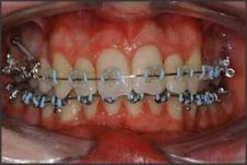
The miniscrews are placed between the upper laterals and the canines or in between the upper canines and the bicuspids for the maxillary arch and in between the laterals and canines for mandibular arch on opposite side for midline correction. (Figure 4)
4. Molar intrusion :[13]
Molar intrusion can be done in the cases where simple molar intrusion is required. Miniscrews can be placed between the roots of the first and second molars. (Figure 5)
5. Molar distalization:
The ideal implant site in palate is between the roots of the first and second premolars.
Risks and Complications of Miniscrews
Complications can arise during miniscrew placement and after orthodontic loading in regard to stability and patient safety.
 | Figure 2
 |
 | Figure 3
|
 | Figure 4
|
 | Figure 5
|
 | Figure 6
|
Complications During Insertion
Trauma to the periodontal ligament or the dental root
Potential complications of root injury include loss of tooth vitality, osteosclerosis, and dentoalveolar ankylosis.[14],[15] Interradicular placement requires proper radiographic planning, including surgical guide with panoramic and periapical radiographs to determine the safest site for miniscrew placement.[16], [17], [18], [19].
Microscrew Slippage
Placement of miniscrews less than 30o from the occlusal plane can increase the risk of slippage. To avoid this, the clinician can initially engage bone with the miniscrew at a more obtuse angle before reducing the angle of insertion after the second or third turn.
Nerve Involvement
Most minor nerve injuries not involving complete tears are transient, with full correction in 6 months.[20] Long-standing sensory aberrations might require pharmacotherapy (corticosteroids), microneurosurgery, grafting, or laser therapy.
Miniscrew bending, fracture, and torsional stress
Increased torsional stress during placement can lead to implant bending or fracture, or produce small cracks in the peri-implant bone, that affect miniscrew stability.[21],[22],[23] Self-drilling miniscrews should be inserted slowly, with minimal pressure, to assure maximum miniscrew-bone contact.
Complications Under Orthodontic Loading
Stationary anchorage failure
According to the literature, the rates of stationary anchorage failure of miniscrews under orthodontic loading vary between 11% and 30%.[24],[25],[26],[27]
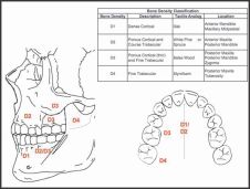
The key determinant for stationary anchorage is bone density.[28],[29],[30]
Stationary anchorage failure is often a result of low bone density due to inadequate cortical thickness. Bone density is classified into 4 groups (D1, D2, D3, and D4) based on Hounsfield units (HU)-an x-ray attenuation unit used in computed tomography scan interpretation to characterize the density of a substance.[31] D1 (>1250 HU) is dense cortical bone primarily found in the anterior mandible and the maxillary midpalatal area.[32] D2 (850-1250 HU) is thick (2 mm), porous cortical bone with coarse trabeculae primarily found in the anterior maxilla and the posterior mandible. D3 (350-850 HU) is thin (1 mm), porous cortical bone with fine trabeculae primarily found in the posterior maxilla with some in the posterior mandible. D4 (150-350 HU) is fine trabecular bone primarily found in the posterior maxilla and the tuberosity region. (Figure 6)
Sevimay et al[33] reported that osseointegrated dental implants placed in D1 and D2 bone showed lower stresses at the implant-bone interface. Placement of miniscrews in D1 and D2 bone might provide greater stationary anchorage under orthodontic loading. Placement of miniscrews in D4 bone is not recommended due to the reported high failure rate.[34],[35],[36].
Miniscrew Migration
Orthodontic miniscrews can remain clinically stable but not absolutely stationary under orthodontic loading. To account for potential migration, the clinician should allow a 2mm safety clearance between the miniscrew and any anatomical structures.[37]
Soft-Tissue Complications
Aphthous ulceration
Minor aphthous ulcerations are self-limiting and resolve within 7 to 10 days without scarring.[38]
Soft tissue inflammation, infection, and periimplantitis
Healthy peri-implant tissue plays an important role as a biologic barrier to bacteria.[39] Tissue inflammation, minor infection, and peri-implantitis can occur after miniscrew placement.[40]
Complications During Removal
Miniscrew fracture
The miniscrew head could fracture from the neck of the shaft during removal. The proper placement technique can minimize the risk of miniscrew fracture during its removal.
Partial osseointegration
Although orthodontic miniscrews achieve stationary anchorage primarily through mechanical retention, they can achieve partial osseointegration after 3 weeks, increasing the difficulty of their removal. The miniscrew typically can be removed without complications a few days after the first attempt of removal.[41]
Conclusion :
The success of this type of absolute anchorage has widened the horizons of the orthodontist, which should be explored to the best possible advantage for treating cases. This should help in providing the aesthetically conscious adult patient orthodontic care which has once compromised or denied altogether due to lack of posterior teeth which serve as anchors during orthodontic treatment.
References
1. Parr GR, Krik L, Toth RW. Titanium: The mystery of implant dentistry. Dental material aspects: J Prosthetic Dent. 1985;54;3:410-14.
2. Carl E. Misch. Contemporary Implant Dentistry.
3. Branemark PI. Osseointegration and its experimental background. J Prosthetic Dent. 1983;50(3):399-410.
4. Martin W, Hafferman M, Ruskin J. Template Fabrication for midpalatal Orthodontic Implant: Technical Note. Int J Oral maxillofacial Implants 2002;17:720-2.
5. Manio BG, Bednar J, Pagein P, Mura P. The spider Screw for skeletal anchorage. JCO 2003;37;2:90-7.
6. Chang J, Lin Y, Jein E, Liou W. A new bone screw for Orthodontic Anchorage. JCO 2003; 37;12:676-81.
7. Kanomi R. Mini implant for Orthodontic Anchorage JCO 1997;31;11:763-7.
8. JCO Interviews :Dr. Junji Sugawara on the skeletal Anchorage System. JCO December 1989;689-96.
9. Clerck HD, Gurinckx V, Siciliano S. The Zygoma Anchorage system. JCO 2002; 36;8:455-9.
10. Glatmaier J, Wehrbein H, Diedrich P. Biodegradable implants for orthodontic anchorage. A preliminary biomechanical study. Journal of Orthodontics 1996;18:465-9.
11. Hashem JK, Mitchell DA. Resorbable implants (Plates and Screws) in orthgnathic surgery. Journal of Orthodontics. 2000;27;5.
12. Kyung H, Park H, Bae S, Sung JH, Kim B. Development of orthodontic micro implant for intraoral anchorage. JCO 2003;37;6:321-8.
13. Sherwood KM, Burch JG, Thompson WJ. Closing Anterior Open bites by intruding molars with titanium miniplates anchorage. Am J Orthod 2002;122:593-600.
14. Asscherickx K, Vannet BV, Wehrbein H, Sabzevar MM. Root repair after injury from miniscrew. Clin Oral Implants Res 2005;16:575-8.
15. Mine K, Kanno Z, Muramato, T, Soma K. Occlusal forces promote periodontal healing of transplanted teeth and prevent dentoalveolar ankylosis: an experimental study in rats. Angle Orthod 2005;75:637-44. 7. Carano A, Velo S, Leone P, Siciliani G. Clinical applications of the miniscrew anchorage system. J Clin Orthod 2005;39:9-24.
16. Morea C, Dominguez GC, Wuo AV, Tortamano A. Surgical guide for optimal positioning of mini-implants. J Clin Orthod 2005;39:317-21.
17. Bae SM, Park HS, Kyung HM, Kwon OW, Sung JH. Clinical application of micro-implant anchorage. J Clin Orthod 2002;36:298-302.
18. Dula K, Mini R, van der Stelt PF, Buser D. The radiographic assessment of implant patients: decision-making criteria. Int J Oral Maxillofac Implants 2001;16:80-9.
19. Suzuki EY, Buranastidporn B. An adjustable surgical guide for miniscrew placement. J Clin Orthod 2005;39:588-90.
20. Ozen T, Orhan K, Gorur I, Ozturk A. Efficacy of low level laser therapy on neurosensory recovery after injury to the inferior alveolar nerve. Head Face Med 2006;2:3.
21. Heidemann W, Gerlach KL, Grobel KH, Kollner HG. Influence of different pilot hole sizes on torque measurements and pullout analysis of osteosynthesis screws. J Craniomaxillofac Surg 1998;26:50-5.
22. Phillips JH, Rahn BA. Comparison of compression and torque measurements of self-tapping and pretapped screws. Plast Reconstr Surg 1989; 83:447-56.
23. Trisi P, Rebaudi A. Progressive bone adaptation of titanium implants during and after orthodontic load in humans. Int J Periodontics Restorative Dent. 2002; 22:31-43.
24. Adell R, Lekholm U, Rockler B, Branemark PI. A 15-year study of osseointegrated implants in the treatment of the edentulous jaw. Int J Oral Surg 1981;10:387-416.
25. Buchter A, Wiechmann D, Koerdt S, Wiesmann HP, Piffko J, Meyer U. Load-related implant reaction of mini-implants used for orthodontic anchorage. Clin Oral Implants Res 2005;16:473-9.
26. Cheng SJ, Tseng IY, Lee JJ, Kok SH. A prospective study of the risk factors associated with failure of mini-implants used for orthodontic anchorage. Int J Oral Maxillofac Implants 2004;19:100-6.
27. Fritz U, Ehmer A, Diedrich P. Clinical suitability of titanium microscrews for orthodontic anchorage-preliminary experiences. J Orofac Orthop 2004;65:410-8.
28. Misch CE. Contemporary implant dentistry. 2nd ed. St Louis: Mosby; 1998.
29. Lee JS, Kim DH, Park YC, Kyung SH, Kim TK. The efficient use of midpalatal miniscrew implants. Angle Orthod 2004;74:711-4.
30. Misch CE. Density of bone: effect on treatment plans, surgical approach, healing, and progressive bone loading. Int J Oral Implantol 1990;6:23-31.
31. Lekholm U, Zarb GA. Patient selection and preparation. In: Branemark PI, Zarb GA, Albrektsson T, editors. Tissue-integrated prostheses: osseointegration in clinical dentistry. Chicago: Quintessence; 1985.
32. Schlegel KA, Kinner F, Schlegel KD. The anatomic basis for palatal implants in orthodontics. Int J Adult Orthod Orthognath Surg 2002;17:133-9.
33. Sevimay M, Turhan F, Kilicarslan MA, Eskitascioglu G. Threedimensional finite element analysis of the effect of different bone quality of stress distribution in an implant-supported crown. J Prosthet Dent 2005;93:227-34.
34. Hutton JE, Heath MR, Chai JY, Harnett J, Jemt T, Johns RB, et al. Factors related to success and failure rates at 3-year followup in a multicenter study of overdentures supported by Branemark implants. Int J Oral Maxillofac Implants 1995;10:33-42.
35. Jaffin RA, Berman CL. The excessive loss of Branemark fixtures in type IV bone: a 5-year analysis. J Periodontol 1991;62:2-4.
36. Suarez D. Miniscrews and the orthodontist: should they be used? Connvention News. Spring 2005 .
37. Liou EJ, Pai BC, Lin JC. Do miniscrews remain stationary under orthodontic forces? Am J Orthod Dentofacial Orthop 2004;126:42-7.
38. Murray LN, McGuinness N, Biagioni P, Hyland P, Lamey PJ. A comparative study of the efficacy of Aphtheal in the management of recurrent minor aphthous ulceration. J Oral Pathol Med 2005;34:413-9.
39. Costa A, Pasta G, Bergamaschi G. Intraoral hard and soft tissue depths for temporary anchorage devices. Semin Orthod 2005;11:10-5.
40. Ohmae M, Saito S, Morohashi T, Seki K, Qu H, Kanomi R, et al. A clinical and histological evaluation of titanium mini-implants as anchors for orthodontic intrusion in the beagle dog. Am J Orthod Dentofacial Orthop 2001;119:489-97.
41. Melsen B, Verna C. Miniscrew implants: the Aarhus anchorage system. Semin Orthod 2005;11:24-31. |