Diagnosis may be defined as identifying disease from an evaluation of the history, signs and symptoms, laboratory tests, and procedures. It is primarily derived from information obtained from the patient's medical and dental histories combined with findings from thorough oral examination. The entire constellation of signs and symptoms associated with disease is taken into consideration before arriving at a diagnosis. In some instances additional information provided by laboratory tests is useful in the over all decision making process.
Traditional periodontal diagnostic parameters used clinically include probing depths, mobility assessment, bleeding on probing, clinical attachment levels, plaque index, and radiographs assessing alveolar bone level. For the most part, findings from traditional diagnostic procedures (e.g., signs of inflammation, deep probing depths, and clinical attachment loss) are related to pathologic processes associated with periodontal infections. Finally, the absence of conventional signs of periodontal disease is strongly related to the presence of a stable, healthy periodontium.
Limitations of traditional periodontal diagnostic techniques:
Clinical or radiological measurements of attachment loss are not precisely accurate and if not carried out very carefully can be misleading.
Full mouth recording is necessary because of the site specific and episodic nature of periodontal disease progression.
Individual susceptibility to periodontitis varies both genetically and over time because of other conditions which may affect susceptibility. Such conditions need to be determined and taken into account.
All clinical diagnostic techniques provide only retrospective information about past disease activity and are unable to diagnose present disease activity.
If regular serial periodontal chartings are to be compared to monitor periodontal progression or if these measurements are required for clinical research purposes then much more accurate diagnostic techniques are necessary.
Commonly used diagnostic procedures have these significant weaknesses. There exists a critical need for objective diagnostic methods to enable clinicians to identify sites in the periodontium having active disease. A great deal of current periodontal research has therefore been directed towards improving this situation. This has been both aimed at improving the accuracy of traditional clinical diagnostic methods and developing alternative methods capable of detecting periodontal disease activity clinically. This article aims to review the advances in diagnostic aids in periodontics.s
Periodontal Probes
The word probe is derived from the Latin word Probo, which means "to test." Periodontal probes are used primarily to detect and measure periodontal pockets and clinical attachment loss[1]. In addition, they are used to locate calculus; measure gingival recession, width of attached gingiva, and size of intraoral lesions; identify tooth and soft-tissue anomalies; locate and measure furcation involvements; and determine mucogingival relationships and bleeding tendencies.
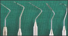
First-Generation (Conventional) Probes
Conventional or manual probes (Fig. 1) do not control for probing pressure and are not suited for automatic data collection.[6] These probes most commonly are used by general dental practitioners as well as periodontists. Invented in 1936 by periodontist Charles H.M. Williams, the Williams' periodontal probe is the prototype or benchmark for all first-generation probes. These probes have a thin stainless steel tip of 13 mm in length and a blunt tip end with a diameter of 1 mm. The graduations on these probes are 1 mm, 2 mm, 3 mm, 5 mm, 7 mm, 8 mm, 9 mm, and 10 mm. (The 4-mm and 6-mm markings are absent to improve visibility and avoid confusion in reading the markings.) The probe tips and handles are enclosed at 130o.
 | Fig. 1
 |
 | Fig. 2
|
The Community Periodontal Index of Treatment Need (CPITN) was designed by Professors George S. Beagrie and Jukka Ainamo in 1978. CPITN probes are recommended for use when screening and monitoring patients with the CPITN index. The index and its probes were first described in World Health Organization's (WHO) Epidemiology, etiology, and prevention of periodontal diseases. Report of a WHO Scientific Group.[9] The FDI World Dental Federation/WHO Joint Working Group 1 has advised the manufacturers of CPITN probes to identify the instruments as CPITN-E (epidemiologic), which have 3.5-mm and 5.5-mm markings, and CPITN-C (clinical), which have 3.5-mm, 5.5-mm, 8.5-mm, and 11.5-mm markings. CPITN probes have thin handles and are lightweight (5 gm). The probes have a ball tip of 0.5 mm, with a black band between 3.5 mm and 5.5 mm, as well as black rings at 8.5 mm and 11.5 mm.
University of North Carolina-15 (UNC-15) probes are color-coded at every millimeter demarcation. They are the preferred probe in clinical research if conventional probes are required.
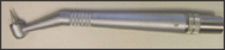
The Naber's probe is used to detect and measure the involvement of furcation areas by the periodontal disease process in multirooted teeth. Naber's probe also is used in the assessment of more complex clinical cases, including those with a restorative treatment. These probes can be color-coded or without demarcation.
Second-Generation (Constant-Pressure) Probes
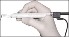
The second-generation instruments are pressure sensitive, allowing for improved standardization of probing pressure. Scientific literature that demonstrated probing pressure should be standardized and not exceed 0.2 N/mm2 led to the development of these probes.Second-generation probes can be used in general dental practices, as well as periodontal practices, and do not require computerization in the operatory [1].
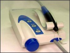
The True Pressure Sensitive (TPS) (Fig. 2) probe is the prototype for second-generation probes. Introduced by Hunter in 1994, these probes have a disposable probing head and a hemispheric probe tip with a diameter of 0.5 mm. A controlled probing pressure of 20 gm is usually applied. These probes have a visual guide and a sliding scale where two indicator lines meet at a specified pressure.
In 1977, Armitage designed a pressure-sensitive probe holder to standardize the insertion pressure and determine how accurate probing pressure of 25 pounds affected the connective-tissue attachment. In 1978, van der Velden devised a pressure-sensitive probe with a cylinder and piston connected to an air-pressure system. Subsequently, it was modified with a displacement transducer for electronic pocket-depth reading.
The electronic pressure-sensitive probe, allowing for control of insertion pressure, was introduced by Polson in 1980. This probe has a handpiece and a control base that allows the examiner to control the probing pressure. The pressure is increased until an audio signal indicates that the preset pressure has been reached. Polson's original design was modified by its initial users: that probe is known as the Yeaple probe, which is used in studies of dentinal hypersensitivity.
 | Fig. 3
|
 | Fig. 4
|
 | Fig. 5
|
 | Fig. 6
|
 | Fig. 7
|
 | Fig. 8
|
 | Fig. 9
|
Third-Generation (Automated) Probes
In spite of the advances in second-generation probes, other sources of errors, such as in reading the probe, recording data, and calculating attachment level, still needed to be addressed. Third-generation probes were developed to help minimize these mistakes by using not only standardized pressure, but also digital readouts of the probes' readings and computer storage of data. This generation includes computer-assisted direct data capture to reduce examiner bias and allows for greater probe precision. These probes require computerization of the dental operatory and can be used by periodontists and academic institutions for research.
The Foster-Miller probe (Foster-Miller, Inc, Waltham, MA) is the prototype of third-generation probes. Devised by Jeffcoat et alin 1986, this probe has controlled probing pressure and automated detection of the cementoenamel junction (CEJ)[6]. The components of the probe are: a pneumatic cylinder, a linear variable differential transducer (LVDT), a force transducer, an accelerator, and a probe tip.
The main mechanism of action of the Foster-Miller probe is by detection of the CEJ. The ball tip moves or glides over the root surface at a controlled speed and preset pressure. Abrupt changes in the acceleration of the probe movement (recorded on a graph) indicate when it meets the CEJ and when it is stopped at the base of the pocket. Under controlled pressure, the probe tip is extended into the pocket and refracted automatically when the base of the pocket is reached. Position and acceleration-time histories are analyzed to determine attachment level and pocket depth. As with all devices, the Foster-Miller probe has advantages and disadvantages. The main advantage is the automatic detection of the CEJ, which is a better landmark than gingival margin, because the position of the gingival margin may change depending on inflammation or recession. The main disadvantage is that it can deem root roughness or root surface irregularities as the CEJ.
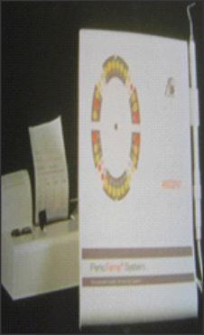
The Florida Probe® (Florida Probe Corp, Gainesville, FL) (Fig. 3) was devised by Gibbs et al in 1988 is probe consists of a probe handpiece and sleeve; a displacement transducer; a foot switch; and a computer interface/personal computer. The hemispheric probe tip has a diameter of 0.45 mm, and the sleeve has a diameter of 0.97 mm. Constant probing pressure of 15 gm is provided by coil springs inside the handpiece. The edge of the sleeve is the reference from which measurements are made, and the probe has Williams' markings; however, actual measurement of the pocket depth is made electronically and transferred automatically to the computer when the foot switch is pressed.
These probes provide a constant probing pressure of 15 gm, which can be overridden when necessary, for accuracy and patient comfort. Each measurement is recorded with potentially 0.2-mm accuracy. Comparison to previous data can be made more quickly and accurately. (The system shows black arrows for changes between 1 mm and 2 mm, and red arrows are used for changes > 2 mm.) Also, there is a chart showing diseased sites, which can be used in patient education. The Florida Probe does have some disadvantages, which include underestimating deep probing depths a lack of tactile sensitivity. Also, clinicians need to be trained to operate these probes.
The Toronto Automated probe, devised by McCulloch and Birek in 1991 at University of Toronto, used the occlusoincisal surface to measure relative clinical attachment levels. The sulcus is probed with a 0.5-mm nickel-titanium wire that is extended under air pressure. It controls angular discrepancies by means of a mercury tilt sensor that limits angulation within ± 30º. This probe has the advantage of an incorporated electronic guidance system to improve precision in probe angulation. It also estimates the biophysical integrity of the dentogingival junction by measuring intrapocket probing velocity. The disadvantages are associated with positioning: it is difficult to measure second and third molars, and patients have to position their heads in the same place to reproduce readings.
The InterProbe™ (The Dental Probe Inc, Glen Allen, VA), also known as the Perio Probe, is a third-generation probe with a flexible probe tip, which curves with the tooth as the probes enter the pocket area. Stainless steel probes push the gingiva away from the tooth, causing pain, whereas the InterProbe gently slides in. This probe produces accurate readings of periodontal pockets with its standardized 15 gm of pressure. The probe's optical encoder handpieces uses constant probing pressure, which provides repeatable measurement of pocket depth and attachment loss.
Fourth-Generation
Fourth-generation refers to three-dimensional (3D) probes. Currently under development, these probes are aimed at recording sequential probe positions along the gingival sulcus. They are an attempt to extend linear probing in a serial manner to take into account the continuous and 3D pocket being examined.
Fifth-Generation
Despite all the advances in earlier generation probes, they remain invasive and, at times, their use can be painful to patients. Plus, with these earlier generation probes, the probe tip usually crosses the junctional epithelium. Fifth-generation probes are being devised to eliminate these disadvantages. Probes are being designed to be 3D and noninvasive: an ultrasound or other device is added to a fourth-generation probe[5]. Fifth-generation probes aim to identify the attachment level without penetrating it.
The only fifth-generation probe available, the UltraSonographic (US) probe (Visual Programs, Inc, Glen Allen, VA) (Fig. 4), uses ultrasound waves to detect, image, and map the upper boundary of the periodontal ligament and its variation over time as an indicator of the presence of periodontal disease. The US probe was devised by Hinders and Companion at the NASA Langley Research Center.[24] This small intraoral probe has an ultrasound beam projection area close enough in size to the width of the periodontal ligament space to give the optimal coupling and small enough to inspect the area between the teeth, while still delivering sufficient signal strength and depth of penetration to image the periodontal ligament space. To probe these structures ultrasonically, a narrow beam of ultrasonic energy is projected down between the tooth and bone from a transducer, which is scanned manually along the gingival margin.
The transducer is mounted at the base of a dual-taper, convergent-divergent coupler to provide an acoustically tapered interface with a throat area on the order of 0.5 mm. This constitutes an active area reduction from the transducer element to the aperture of 20:1. Such a reduction is mandated by the geometry and the very small window afforded by the gingival margin. An added virtue of attaining this small a tip size is the ability of the ultrasonic probe to help the clinician examine the area between the teeth, which is where periodontal disease is most likely to occur.
The ultrasound transducer is mounted in the probe-tip shell, which also incorporates a slight flow of water to ensure good coupling of the ultrasonic energy to the tissues. The couplet water can come either from a suspended intravenous-type sterile bag or plumbed from the dental-unit water source. The focused ultrasonic beam is transmitted into the pocket in the same orientation as the insertion of a manual probe Then, the probe is moved along the gingival margin, so the two-dimensional graphical output corresponds to the results a clinician gets from "walking the sulcus" with a manual probe. However, ultrasound gives more information because secondary echoes are recorded from tissue features at various depths. It appears likely that the technique also will be able to provide information on the condition of the gingival tissue and the quality and extent of the epithelial attachment to the tooth surface. This may supply valuable data to aid the clinician in the diagnosis and treatment charting of these diseases.
Nonperiodontal Probes
Calculus Detection
Calculus detection probes detect subgingival calculus by means of audio readings and are reported to increase chances of subgingival calculus detection[7]. Currently, the DetecTar probe (DENTPLY Professional, Des Moines, IL) (Fig. 5) is the only calculus detection probe on the market. This device has a lightweight, well-balanced handpiece, which can be autoclaved, and it produces an audible beep to signify calculus detection (beep function can be disengaged). This probe may augment standard methods of calculus detection; however, it is expensive and the handpiece is bulkier than a standard periodontal probe. The probe has a short waterline hookup, which may prevent ergonomic placement of the unit, and it does not have a published waterline treatment protocol. As with many automated probes, there is potential for false positives and false negatives; therefore, further research is required.
Temperature Sensitive Probe
The Periotemp® Probe (Abiodent Inc, Danvers, MA) (Fig. 6) is a temperature-sensitive probe, which reportedly detects early inflammatory changes in the gingival tissues by measuring temperature variations in these tissues[4]. The Periotemp Probe detects pocket temperature differences of 0.1oC from a referenced subgingival temperature.This probe has two light indicating diodes: red-emitting diode, which indicates higher temperature, denoting risk is twice as likely for future attachment loss; and green-emitting diode, which indicates a lower temperature, indicating lower risk. This probe can detect initial inflammatory changes; therefore, treatment can be initiated at an early stage. However, the presence of surface cooling caused by breath airflow may further complicate the determination of even a normal temperature distribution.
Hallimeter
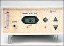
Halitosis or Oral malodor is most often caused by Gram -ve anaerobic bacteria which degrades the proteins and produce volatile sulphur compounds[7]. These sulphur compounds can be detected by gas chromatography or the recently Hallimeter (Interscan)(Fig.7) by Rosenberg et al in 1991.The Hallimeter is compact ,easy to use and allows chairside testing. Its performance lacks specificity in the analysis of the different components of mouth air in comparison to the gold standard gas chromatography with flame photometric detection.( Tonzetich et al 1995).
Diamond Probe
This is a recently developed commercially available instrument developed by the Diamond General Development Corporation, Ann Arbour USA(Fig. 8). It has been designed so that it combines the features of a periodontal probe with the detection of volatile sulphur compounds in the periodontal pocket.
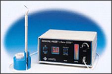
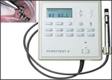
Periotest
Tooth mobility is a clinical expression of periodontitis.The Periotest (Fig. 9) is one of the current systems to assess tooth mobility.It utilizes dynamic forces of short duration of low millisecond range.It evaluates the damping characteristics of the tooth,the device consists of a hand piece that is connected to a unit via a cable.Inside the hand piece a metal rod is accelerated until it reaches its nominal speed and contacts the tooth.The tooth is slightly deflected and the rod is decelerated.Using the measured contact times in milliseconds the Periotest values are calculated[7].
The following ranges are recorded.
-8 to +9-clinically firm tooth
10-19 -Palpable mobility
20-29 -Visible mobility
30-50 -Mobility in response to lip and tongue movements.
Errors may occur due to variation in duration,point of application,mode of application,manner and duration and time of forces,or due to instability variation,slippage of device etc.
Conclusion:
Although there are many potential clinical diagnostic methods for assessing periodontal disease activity and progression, still numerous features hamper the ability to use them as diagnostic methods of proven utility. There is still a lack of a proven gold standard of disease progression. After all these years of intensive research we look forward for the betterment of the diagnostic aids to measure present disease activity.
References
1. Arthur F.H Periodontal probing Crit. Rev. Oral Biol. Med. 1997;8:336-356.
2. Badersten A, Nilveus R, Egelberg J. Reproducibility of probing attachment level measurements. J Clin Periodontol 1984;11:475-485.
3. Clark WB, Yang MC, Mangusson I. Measuring clinical attachment: reproducibility of relative measurements with an electronic probe. J Periodontol 1992;63:831-838.
4. Haffajee AD, Socransky SS, Goodson JM. Subgingival temperature. I. Relation to baseline clinical parameters. J Clin Periodontol 1992: 19: 401-408.
5. Jidog et al Ultrasonic periodontal probing based on the dynamic wavelength fingerprint. EURASIP Journal on applied signal Processing 2005;7:1137-1146.
6. Lang , Lindhe. Clinical Periodontology and Implant Dentistry 5th ed
7. Newman MG, Takei HH, Carranza FA, Klokkevold PR Carranza's Clinical Periodontology 10th ed.
8. N. Surathu Current trends in periodontal diagnosis, disease recognition and management Proceedings of the 5th Asian Pacific Society of Periodontology Meeting Cebu, the Philippines 2003
9. Pihlstrom Periodontal risk assessment, diagnosis and treatment planning Periodontology 2000, Vol. 25, 2001, 37-58 |