Introduction
Modern dentistry is evolving at a rapid rate with the advent of new materials and technologies. One suchtechnique which has undergone explosive amelioration is the use of lasers in dental practice. After their introduction in the 1960s, lasers have had diverse applicability in clinical dentistry.[1] Amongst their many usesis laser-assisted tooth bleaching.
Tooth bleaching is becoming an increasing sought after treatment modality as patients are exceedingly concerned about aesthetics. Tooth discolouration may influence an individual to have a compromised self image and thus, bleaching treatments provide both aesthetic as well as psychological benefits.[2]
It was only in 1996 that laser-assisted bleaching was officially started with the approval of Ion Laser Technology's argon and carbon dioxide lasers by the FDA.[3] Power bleaching using lasers was introduced in order to accelerate and enhance the bleaching process.[4] Laser bleaching is considered to be more efficacious as one can obtain controlled temperature elevation of the bleaching agent, and by photochemical activationprovide a higher overall yield of intrinsic radicals causingtheir better absorption into the dental hard tissues. This collectively minimises adverse pulpal reactions by reduced thermal activation of the bleaching agents.
Now-a-days KTP-L (potassium titanyl phosphate laser), argon and diode lasers are commonly used for in-office power bleaching.The versatility of the diode laser is one of its most distinguishing characteristics. They have a multitude of applicability which includes soft tissue surgery, periodontal therapy, implantology, endodontics and esthetic dentistry.[5] Being a "soft" or "cold laser" it has also been employed for use in Low Level Laser Therapies (LLLT). The diode laser when used in bleaching procedures has been found to cause a temperature increase in pulp chamber below the critical temperature of 5.5oC, regarded as the threshold to prevent irreversible pulpal change.[6] A preclinical investigation using diode laser bleaching has concluded that it is a valuable energy source, and is simple as well as effective.[7]
The following case report describes the clinical application of the diode laser forvital tooth bleaching.
Case Report
A 26-year old female patient visited our dental clinic with the chief complaint of generalized discolouration of teeth and wanted her teeth bleached to make them cosmetically desirable. She had noted this discolouration since the last 3 years during which she had undergone orthodontic treatment. On clinical examination we observed that all the teeth had a yellowish discolouration. (Fig. 1)
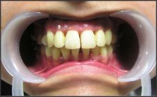 | Fig. 1. Pre-operative photograph showing generalised discolouration of teeth
 |
After explaining the various treatment options and the procedure to be undertaken, informed consent was taken from the patient.
The first step was to carry out shade matching of the teeth in order to set a bench mark for later comparison. This was followed by polishing the teeth using a pumice paste in order to remove plaque. Cotton wool isolation was used to maintain a dry field and a cheek tractor was used for better accessibility. The patient was made to wear protective eye wear all through the procedure. Vitamin E was kept available along with cotton tip applicators as this cures any irritation in cases where the bleaching gel comes in touch with soft tissues.
No local anaesthesia or pre-emptive analgesics were administered during treatment. Gingival protection was provided using a resin barrier (OpalDam, Ultradent) which was extended onto the cervical margin of the teeth by approximately 1mm.
The bleaching gel (JWhite Power Bleaching Gel, Heydent) was then prepared as per the manufacturer's instructions and applied onto the teeth using an applicator tip. The gel was first applied on the maxillary anterior teeth and gradually advanced towards the premolars bilaterally. The gel was then applied on the mandibular teeth in the same sequence. (Fig. 2)
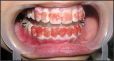 | Fig. 2. Photograph showing application of bleaching gel on teeth and resin barrier protection on adjacent gingiva.
|
A diode laser (Fotona XD-2) with a special bleaching hand piece (R24-B) was used for irradiation. Each tooth was exposed to the laser for 30 seconds in the same sequence as that of gel application. The laser tip was used in non-contact mode and was constantly rotated in order to prevent prolonged focal application. The average power setting of the laser was maintained at 4 Watts.
After all the teeth had been irradiated the bleaching gel was aspirated and any remaining gel was removed using cotton pellets. The patient was then made to rinse and the teeth were dried. Thereafter the tooth colour was compared to the original recording.A second exposure was then done in order to achieve the desirable result. (Fig. 3)
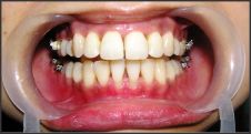 | Fig. 3. Post-operative photograph after diode laser assisted tooth bleaching showing improvement in the teeth shades.
|
The patient was requested to come to the clinic after a period of 10 days to check for any reversal in the tooth colour. The patient was also asked to report to the clinic in case of any discomfort.
The patient reported back to the clinic without any history of sensitivity or irritation. On clinical examination no setback in the tooth colour was noted.
Discussion
The etiology of tooth discolouration is multivarious. Stains on teeth can be classified as either extrinsic or intrinsic. Extrinsic stains are usually seen in inaccessible areas and may result from plaque, beverages, dietary precipitates, medications or iron supplements. Some of the causes for intrinsic staining include trauma, diseases (like erythroblastosis fetalis, porphyria, amelogenesis imperfecta), medications (eg. tetracycline), aging, etc.
Numerous techniques for teeth bleaching have been introduced in dentistry and continually reformed. Diverse numbers of oxidizing agents have been employed in tooth whitening and found to cause adverse effects on dental hard tissues and are advised to be used with caution.[8],[9] On the other hand, laser tooth bleaching causes less change in microhardness of enamel and does not result in much temperature elevation.[10] Another factor in favour of laser bleaching is the amount of time required to achieve desirable results. In today's society, where time is of essence, laser bleaching would be preferred over other techniques as 60-90 minutes would produce a favourable outcome.
The diode laser we used is provided with a specialized handpiece for laser tooth bleaching. This allows a laser spot-size of 6mm and the appropriate wattage, preventing any thermal insult on the pulp. The laser is easy to manipulate and extremely versatile.
The resin barrier protection (OpalDam, Ultradent) we used is a light-reflective, passively adhesive, light cure resin. It is a methacrylate based resin which maintains adequate strength where needed and at the same time can be removed easily from embrasures and undercuts.
We utilized JWhite Power Bleaching gel which is prepared by mixing hydrogen peroxide with JWhite powder as per the manufactures instructions. Use of 38% hydrogen peroxide activated by diode laser has been found to result in improved enamel luminosity using digital photometry.[11] Also, unlike other bleaching agents peroxide gels have a basic pH and this prevents any surface alterations or demineralization of the tooth surface.
The diode laser provides quick and effective results when used to activate the bleaching gel. An in vitro study concluded that diode laser activation of bleaching agent presented significantly better results as compared to the agent being used alone or when combined with LED source.[12] An added advantage is the reduced contact time of the bleaching agent with the teeth and this minimizes the chances of sensitivity or irritation.
Conclusion
In conclusion, we would like to state that diode laser tooth bleaching is a viable treatment option, as it provides fast and effective bleaching without causing any thermal damage. The reduced time frame when utilizing power tooth bleaching will result in better patient compliance and satisfaction. Further investigation to prove the efficacy of laser tooth bleaching should be undertaken.
References
1. Goldman L, Hornby P, Meyer R and Goldman B (1964). Impact of the laser on dental caries. Nature,203: 417.
2. Dunn J. Dentist prescribed home bleaching: current status. Compend Contin Educ Dent. 1998;19(8):760-4.
3. Greenwall L, editor. Bleaching techniques in restorative dentistry - An illustrated guide. Martin Dunitz Ltd; 2001.
4. Sun G (2000). The role of lasers in cosmetic dentistry. Dental Clinics of North America. 44(4): 831-850.
5. Samo Pirnat. Versatality of an 810 nm diode laser in dentistry : An overview. J Laser Health Acad. 2007. Available from: http://www.laserandhealth.com.
6. Sulieman M, Rees JS, Addy M. Surface and pulp chamber temperature rises during tooth bleaching using a diode laser: a study in vitro. Br Dent J 2006;200:631-63.
7. Dostalvo T, Jelinkiva H, Housova D, Sulc J, Nemec M, Miyagi M, Brugnera Junior A, Zanin F. Diode laser-activated bleaching. Braz Dent J 2004;15:S13-18.
8. Rotstein I, Dankner E, Goldman A, Heling I, Stabholz A, Zalkind M. Histochemical analysis of dental hard tissues following bleacing. J Endod. 1996;22(1):23-26.
9. Lewinstein I, Hirschfeld Z, Stabholz A, Rotstein I. Effect of hydrogen peroxide and sodium perborate on the microhardness of human enamel and dentin. J Endod. 1994;20(1):61-63.
10. Gomes MN, Francci C, Medeiros IS, Salgado NR, Rilil H, Marasca JM, Muench A. Effect of light irradiation on tooth whitening: enamel microhardness and colour change. J Esthet Restor Dent. 2009;21:387-398.
11. Kafas P, Theodoridis M, Dionysopoulos D, Andreou I and Dabarakis N. Diode Laser Tooth Whitening Improved Enamel Luminosity: A Case of Digital Photometry. Research Journal of Medical Sciences. 2008;2:182-184.
12. Wetter NU, Barroso MC, Pelino JE. Dental bleaching efficacy with diode laser and LED irradiation: an in vitro study. Lasers Surg Med. 2004;35(4):254-8. |