Introduction
Turner's hypoplasia usually manifests as a portion of missing or diminished enamel on permanent teeth. Unlike other abnormalities which affect the large number of teeth, Turner's hypoplasia affects only tooth in the oral cavity and it is referred to as a Turner's tooth. If involves single tooth, most likely cause is traumatic injuries leading to primary tooth being pushed into the developing tooth underneath it, infection from fractured deciduous tooth, iatrogenic from inelegant deciduous tooth extraction, bruising from local facial trauma consequently affects the formation of enamel. The affect to trauma is more pronounced if it occurs prior to 3rd year of life2. Injuries occurring after this time are less likely to cause enamel defects since enamel is already calcified. Because of the topographic relationship of the primary teeth to the permanent tooth germ, the most likely affected area on the permanent tooth is the facial surface (the site closer to lips and cheek).[1]
In considering treatment options (extractions, dentin bonding, composite restoration, cast metal restorations) a number of factors need to be considered including:
Number of teeth affected
Location of defects (crown tip, mid crown)
Importance of teeth affected
Severity of defects (coronal and root)
Patient's expectation and resources
Operator's knowledge, skills and resources
Case Report
An 18 year old patient reported to department of Conservative Dentistry and Endodontics with chief complaint of malformed right upper front tooth since childhood. (Fig - 1) Patient gave history of loss of primary incisors due to trauma at the age of 2 years. Clinical examination showed the presence of moderate to severe yellowish brown discoloration with hypoplastic enamel of right lateral incisor. Right lateral incisor exhibited no mobility with vitality test positive .Patient wanted immediate esthetic restoration of malformed tooth due to her personal reasons. So treatment using adhesive resin was preferred as it could be done in single sitting and gave immediate esthetic results. Composite veneering was done on labial surface of right lateral incisor.(Fig - 2)
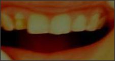 | Fig 1 :Turner' Tooth
 |
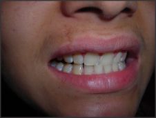 | Fig 2 : Composite veneering
|
After anesthesia was administered, rubber dam isolation was accomplished. Labial reduction was done upto 1mm depth. .the total etch technique was utilized due to its ability to minimize the potential of microleakage and enhance bond strength .The tooth surface was etched for 15 sec with phosphoric acid, rinsed for 5 sec, gently dried for 5 sec, and lightly air thinned to avoid desiccation. The surface was remoistened with water and a hydrophilic adhesive agent (Prime and Bond NT, Dentsply /Caulk, Milford, DE) was applied with a disposable brush. Using continuous motion ,the excess adhesive was removed with a dry micro brush applicator and light cured for 20 sec. Composite resin was applied in increments on the labial surface and each increment was cured for 40 sec. Once the composite was polymerized, a long needle shaped finishing bur was used to finish the labial surface according to the proper anatomic contours of the facial aspect of anterior tooth. Initial finishing was achieved with a 12 fluted, needle shaped bur to replicate natural form and texture. A smooth surface was achieved by following a sequential increase in number of flutes. The facial surface was subsequently polished to a high luster using aluminium oxide discs, prepolish and high shine silicon rubber points(Indentoflex Kerr /Sybron,Orange CA:Astropol,Invoclar Vivadent, Amherst, NY).To impart a high luster and reflectivity on the tooth, The final polishing was accomplished with composite polishing paste and goat hair brushes applied at conventional speed. After polishing procedure was completed ,a final 2 min post curing was performed to improve the degree of conversion and ensure surface hardness.
Discussion
In the present case report it was seen that traumatic injuries to primary predecessors had lead to developmental disturbances in their successor. The permanent tooth which erupted crown defect like defective enamel formation. In the case of amelogenesis, it is not different cells doing different jobs, but the same cells at different stages of maturation doing the different jobs. First they lay down the organic matrix and then they lay down the hydroxyapatite crystals within this matrix and finally they become quiescent and vestigial.[3] Once the cells have matured from one phase and moved to the next, they cannot go back and fix any defects. Anything that disrupts the delicate ameloblasts during enamel production will result in defective enamel which may be very porous and weak. This defective enamel is often present at eruptionbut will soon be lost to abrasive forces. This leaves an area of exposed dentin and rough margins to the surrounding enamel. In some instances the enamel does not form at all and so is missing as soon as the tooth erupts.
Brownish discoloration occurs due to disturbance in ameloblastic layer leading to defective matrix formation caused by traumatic injuries but the stretched inner enamel epithelium continue to induce the differentiation of new odontoblast . Under the defective or missing enamel is dentin, which eventually makes up the bulk of the tooth. It is about as hard as bone but much softer than enamel. It is pale yellow in color, compared to the stark white of normal enamel. In the present case tooth was unaffected by carious lesion ,so it was possible to save the tooth with turner's hypoplasia with minimum intervention and at six months recall visit the patient was asymptomatic ,had maintained esthetics and stability.(Fig - 3)
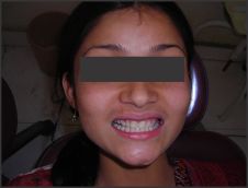 | Fig 3 : Improved esthetics
|
References
1. Kalra N.Sequlae of neglected pulpal infections of deciduous molars.Endodontology 1994;6:19-23
2. Esthetic and endosurgical management of turner's hypoplasia;a sequlaeof trauma to developing tooth germ.J Indian Soc Pedod Prevent Dent,S121-124Supplement-2008.
3. Enamel hypoplasia.www.toothvet.ca.January 2010
4. Brook AH, Fearne JM, Smith J: Environmental causes of enamel defects. Ciba Foundation Symposium 205:212-221,1997.
5. Koch MJ, Garcia-Godoy F: The clinical performance of laboratory-fabricated crowns placed on first permanent molars with developmental defects. JADA 131:1285-1290, 2000.
6. Li RW: Adhesive solutions: report of a case using multiple adhesive techniques in the management of enamel hypoplasia. Dent Update 26:277-287, 1999.
7. Murray JJ, Shaw L: Classification and prevalence of enamel opacities in the human deciduous and permanent dentitions. Arch Oral Biol 24:7-13, 1979.
8. Quinonez R., Hoover R, Wright JT: Transitional anterior esthetic restorations for patients with enamel defects. PediatrDent 22(1):65-67, 2000.
9. Rugg-Gunn AJ, Al Mohammadi SM, Butler TJ: Malnutrition and developmental defects of enamel in 2- to 6-year-old Saudi boys. Caries Res 32:181-192, 1998.
10. Seow WK: Enamel hypoplasia in the primary dentition: a review. ASDC J Dent Child 58:441-452, 1991.
11. Silberman SL, Trubman A, Duncan WK, Meydrech EF: A simplified hypoplasia index. J Public Health Dent 50:282-284, 1990.
12. Slayton, R.L., Warren, J.J., Kanellis, M.J., Levy, S.M. and Islam, M. Prevalence of enamel hypoplasia and isolated opacities in the primary dentition. Pediatric Dentistry 23:32-36, 2001.
13. Witkop CJ, Jr.: Amelogenesis imperfecta, dentinogenesis imperfecta and dentin dysplasia revisited: problems in classification. J Oral Pathol 17:547-553, 1988. |