Discussion:
Class-II Div1 malocclusion i.e. the anterior positioning of upper 1st molars in relation to the lower 1st molars is a very common problem in the children today. Though the genetic component does dominate the incidence,[4] the lifestyle changes in the urban population has given a new dimension to the same. The recent evidences show an increased incidence of nursing bottle caries, rampant caries, early exfoliation of deciduous teeth especially in the upper arch, thus leading to mesial migration of upper 1st molar thereby developing a Class-II Div1 malocclusion . A skeletal Class-II Div1 malocclusion is indeed easily managed in the mixed dentition stage with functional appliances but a developing dental Class-II Div1 malocclusion is somewhat mis understood & mismanaged by general practioners. An interceptive treatment is often not suggested, rather the patient is advised to wait till the eruption of full compliment of permanent teeth. Intercepting these cases early by upper molar distallisation before the eruption of 2nd molars gives the advantage of bringing the molar to its naturally destined position thereby creating room for the normal eruption of anterior teeth & a class-I intercuspation of the upper & lower 1st molars.[5],[6]
Case Report 1:
The patient reported to the clinic at the age of 8 yrs with missing upper right & left deciduous 2nd molars. On examination it was found that the patient had a dental class-II molar relation. Both upper first permanent molars had migrated mesially and were rotated mesiopalatolly, leaving little space for the erupting 2nd bicuspids. The consequences of this developing malocclusion were discussed with the parents of the patient and it was decided to intercept the developing malocclusion. Patient's impressions were made & a pendulum appliance was fabricated to distallize the maxillary 1st permanent molars. The pendulum appliance was activated to deliver distally directed forces on the upper 1st permanent molars thus pushing them back into the maxillary arch in order to create a sufficient space in the maxillary arch for the erupting 2nd bicuspids. The appliance was kept in place for a period of eight months in which time the molars were sufficiently distallized to create adequate space for the erupting premolars And a super Cl-I molar relation was achieved.
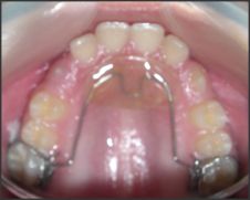
At the end of eight months the maxillary molars had been pushed back in Class-I relationship with respect to lower 1st permanent molars. After the correction had been achieved the appliance was removed and a Nance button was cemented in the upper arch & a lingual arch was cemented on the lower arch till the eruption of the cuspids & bicuspids was complete. The developing malocclusion was thus successfully intercepted and the treatment ended without the need for braces or active orthodontic treatment.
 |
 |
 |
|
 |
|
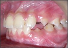
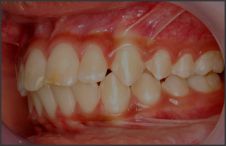
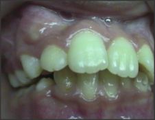
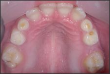
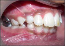
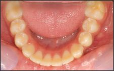
 | Fig:1 ( Before Interception) showing premature loss of maxillary deciduous molars & mesially migrated & severely rotated upper right first permanent molar and Cl-II molar relationship of the upper and lower dental arches.
|
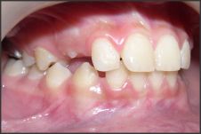
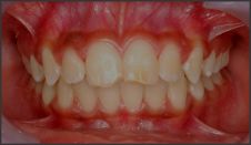
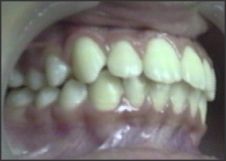
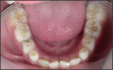
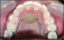
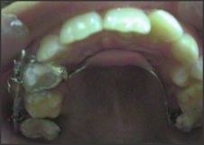
 | Fig:2 (Post-interception) showing the corrected upper molars which have been derotated & distallized . The correction is being maintained by the use of a Nance's button. Note: adequate space has been created for the eruption of the subsequent permanent te
|
Case Report 2:
The patient reported to the clinic at the age of 9 yrs. with a developing Class-II Div1 subdivision on left side with inadequate space for the eruption of maxillary canines. The treatment planning included the distallization of the maxillary 1st permanent molars to create space for the normal eruption of cuspids & premolars and to achieve a Class-I molar relationship. A pendulum appliance was fabricated & cemented on the maxillary arch. The appliance was kept in place for a period of five months in which time an adequate distallization of the maxillary molars was achieved to bring the molars in a class- I relationship, as well as an adequate space was achieved for the subsequent eruption of all maxillary cuspids & bicuspids.
Since the patient also had proclined upper anterior teeth and an increased overjet, the corrective orthodontic treatment with braces was followed to retract the upper anterior teeth in Class-I occlusion. And we were able to treate the case nonextraction thus saving the upper 1st bicuspids from being extracted if the case was delayed till after the eruption of 2nd permanent molars.
 |
|
 |
|
 |
|
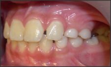
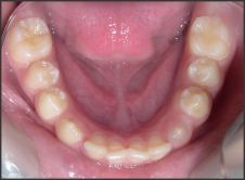
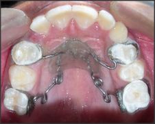
 | Fig:3 (Before Interception) showing a developing Cl-II Div1 malocclussion with an increased overjet , proclined upper/lower anterior teeth & buccally blocked out right canine. The Upper occlussal view shows retained Left & right deciduous canines and inad
|
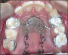
 | Fig: 4 showing the amount of activation built into the pendulum appliance and the pendulum appliance in place exerting a distallizing force on the molars.
|
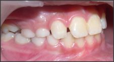
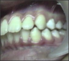
 | Fig: 5 Post-treatment photographs of the patient showing a good Cl-I intercuspation on the right & left side and a normal overjet & overbite.
|
Case Report 3:
A patient aged 8 yrs. reported to the clinic with a history of premature extraction of upper right deciduous canine & first molar and subsequent loss of space by mesial migration of permanent 1st premolar into the extraction space of deciduous canine so that there was no space left for the eruption of upper right permanent cuspid. The patient presented with a developing Class-II Div1 Subdivision on the right side. It was decided to intercept the developing malocclusion without any further delay. The upper right molar was planned to be distallized with the Jones jig appliance. The appliance was cemented in position with bands on upper right first premolar and permanent right first molar.
 | Fig: 6 showing a labially blocked out upper right canine with no space for it's eruption in the arch and distallization of upper right 1st molar with a jones jig appliance.
|
 | Fig:7 Showing post-treatment results with proper alignment of the blocked out upper right canine into the arch after successful distalization of upper right 1st molar and a good Cl-I intercuspation in the posterior segment.
|
The distallization of maxillary right 1st molar took place in a period of eight months creating enough room for the maxillary right cuspid to erupt in the right place as well as a Class-I molar relationship was also achieved on the right side. A Nance button was cemented in place till the eruption of all permanent teeth was complete. The interceptive treatment was followed by the corrective orthodontic treatment for minor correction of alignment after a gap of one year when all the permanent teeth had erupted.
Conclusion:
The interception of a developing Class-II Div1 malocclussion of dental origin due to the mesial migration of upper 1st permanent molars in the late mixed dentition stage before the eruption of permanent 2nd molars has much greater chances of success as less resistance is experienced by the distalizing 1st molars in the absence of the second molars. This gives the clinician enough room in the maxilla to attain an ideal Class-I molar relationship of teeth as well as avoid the extraction of upper 1st bicuspids which is the line of treatment for the correction of increased overjet in a full blown Class-II Div1 malocclusion of dental origin. The amount of distal movement of the maxillary first molars is significantly greater and the anchorage loss is significantly less before the eruption of the second molars. The time taken for molar distalization is also significantly shorter before the eruption of second molars.[7] The reason why it is more effective to move the maxillary first molars distally before the second molars have erupted is, of course, that there is one more tooth, and thus, a larger area of root surface to be moved when the second molars have erupted. Thus, if one has to decide whether to move the maxillary molars distally in the mixed dentition or in the permanent dentition, it is always an advantage to make this intervention at an early age.
References:
1. Davenport IB. The significance of the natural form and arrangement of the dental arches of man, with a consideration of the changes which occur as a result of their artificial derangement by filling or by the extraction of teeth. Dent Cosmos 1887;29:413-39.
2. Hoffding J, Kisling E. Premature loss of primary teeth, part I: its overall effect on occlusion and space in the permanent dentition. ASDC J Dent Child 1978;45(4):279-83.
3. Kimmel NA, Gellin ME, Bohannan HM, and Kaplan AL: Ectopic eruption of maxillary first permanent molars in different areas of the United States. J Dent Child1982, 49: 294-299,
4. Kathryn FS , Thomas JK, Eunice W: Influence of heredity in the etiology of malocclusion. American Journal of Orthodontics Volume 42, Issue 2, February 1956, Pages 125-141
5. Gianelly AA. Distal movement of the maxillary molars. Am J Orthod Dentofac Orthop. 1998;114:66-72.
6. Ghosh, J. and Nanda, R. Evaluation of an intraoral maxillary molar distalization technique, American Journal of Orthodontics and Dentofacial Orthopedics, 1996, 110, 639-646.
7. Papadopoulos MA, Mavropoulos A, Karamouzos A. Cephalometric changes following simultaneous first and second maxillary molar distalization using a non-compliance intraoral appliance. J Orofac Orthop. 2004;65:123-136. |