Introduction
Adhesive dentistry has revolutionised restorative dental practice during the last 30 years. Improved adhesive materials have made resin based composite restorations more reliable and long lasting[1]. With continuous improvement in material science, the indications for composites have progressively shifted from the anterior to the posterior segments of the dental arches[2]. Clinicians are encouraged to place resin composites in increments to ensure complete polymerization of large restorations[3].
It has been hypothesized that dentin surface can absorb salivary glycoprotein, rendering the surface less favourable to bonding[4]. The so called one bottle systems have gained broad acceptance based on the hypothesis that the hydrophilic adhesive solutions in particular the acetone or ethanol based products, may displace or diffuse through a saliva film to reach the underlying hydroxyapitite or collagen as a condition for firm bonding after polymerization.[5]
The purpose of this study was to evaluate (1) The effect of salivary contamination before and after curing of one step self etching bonding agents , (2) efficacy of decontamination method on shear bond strength, (3) effect of reapplication of bonding agent on shear bond strength.
Methods And Materials.
Bonding agents were applied according to manufacturer's instructions. All teeth were restored with Filtek Z250 (3M ESPE, St Paul, MN, USA). A total of one hundred and five freshly extracted non carious permanent intact maxillary premolar teeth were selected for this study. Following extraction, teeth were cleaned by removing any remaining soft tissue tags and then stored in 0.5% thymol solution along with distilled water at 4oC.
Sample Preparation
Teeth were mounted in a cylindrical mould filled with self curing denture base resin upto cervical region (Fig 1). Occlusal surfaces of teeth were reduced along the long axis of the tooth on a water cooled, model trimming wheel to create a flat dentin surface (Fig 2). Immediately prior to the bonding procedure, fresh human saliva was collected from a single individual, The specimens were randomly divided into three major groups (Group A, B, C) of 45 samples each according to salivary contamination protocol. Group A (salivary contamination was done before curing of bonding agent),Group B (salivary contamination was done after curing of bonding agents) and Group C served as control in which no salivary contamination was done. These groups were further subdivided into three subgroups (I, II, III ) of 15 samples each according to bonding agents used which were Xeno III, Adper SE Plus, Clearfil S3. These subgroups were further divided into three mini groups (A1, A2, A3, B1, B2, B3) of 5 samples each, according to decontamination method undertaken. Control Group was divided into three subgroups of 5 samples each (I, II, III) according to bonding agents used, in which no contamination or decontamination protocol was undertaken.
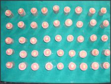 | Fig 1 Mounted Samples
 |
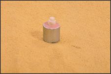 | Fig 2 Occlusal Surface Ground
|
Methodology:
Control Group (C). Adhesive was applied to dentin surface according to the manufacturer's instructions and cured. A tube of an internal diameter 5mm was cut into 2 mm long pieces using a measuring gauge and BP Knife blade to ensure parallel ends. These tubes were placed on the teeth. Resin composite Filtek Z250 (3M ESPE) was carried into the tubes with the help of composite placing instrument, and was cured with the curing light for 20 seconds. The tubes were carefully removed with the scalpel blade prior to testing. All specimens were stored in distilled water at 370C for 24 hours.
Group A Bonding procedure was carried out as in control ; however fresh saliva was applied with an applicator tip to dentin bonded layer before light curing of bonding agent and was left undisturbed for 15 seconds.
Subgroups were as follows:
SubGroups
A1:- Saliva was removed with a gentle air blast. Adhesive was not reapplied. The adhesive was light cured for 20 seconds.
A2 :- Saliva was rinsed with an air water spray from an air water syringe for 10 seconds and then dried with a gentle air blast. The adhesive was then light cured for 20 seconds.
A3:- Saliva was rinsed dried as in A2, a single coat of adhesive was reapplied to dentin surface and light cured for 20 seconds.
Group B
Bonding procedure was carried out as in control; however surface was contaminated with fresh saliva after curing the adhesive. The saliva was left undisturbed for 15 seconds and decontamination protocol was followed as described earlier leading to formation of subgroup B1, subgroup B2 and subgroup B3.
Specimens in each group were tested in shear mode using chisel shaped rod in an universal testing Machine (Model 5582, Instron). A shear force was applied to each specimen at a crosshead speed of 1 mm/minute (Fig 3). The microshear bond strength was calculated by dividing maximum load at failure by cross sectional surface area of the bonded surface using the formula SHEAR STRESS=LOAD (N)/AREA(mm[2])
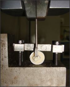 | Fig 3 Applicaiton Of Shear Force
|
Stress was recorded in Mega Pascal(MPa). Load was recorded in Newton (N). Area was calculated by using formula r[2] .Radius of the tube was 2.5mm, so by applying the formula calculated area was 19.625 mm[2] for each specimen.
Stastistical Analysis
The data was subjected to unpaired t Test to make comparison among the groups (Fig 4).
 | Fig 4 Statistical Analysis
|
Results
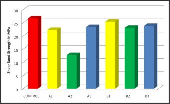
Table 1 summarizes the mean shear bond strengths in MPa and standard deviation for the different groups and subgroups (Mean ±SD). Statistically significant difference between Group A, GroupB and control were observed.
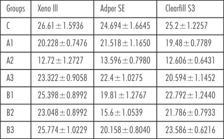 | Table 1 Summarizes the mean shear bond strengths in MPa and Standard Deviation for the different groups and subgroups (Mean ±SD)
|
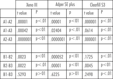 | Table 2: A comparative evaluation within the same group amongst the subgroups
|
When a intergroup comparison of the shear bond strength values between the subgroups of all the three adhesives, significant difference was observed for A1-B1, A2-B2 and A3-B3.
By using unpaired t test , a significant level of difference between A1-A2, A2-A3, A1-A3 and B1-B2,B2-B3, for the three material Xeno III, Adper SE plus ,Clearfill S3, was observed among subgroups. No significant difference was observed among subgroup B1-B3 for Adper SE plus and Xeno III.
Discussion
It has been an established fact for decades now that bonding protocol needs proper isolation of the preparation for prevention from contamination of any kind. Indeed moisture and in particular saliva can affect the quality of the bond, leading to micro leakage or even bond failure. This would result in post operative sensitivity, discoloration, recurrent caries and even loss of restoration[6]. Protein contamination of the operating field from inadvertent contact with blood or saliva is a frequent problem in dentistry when rubber dam isolation is not practised.[7]
Only about 17% of professionals routinely use the rubber dam. In this context, the contamination of the operating field is a recurrent reality that emphasizes the value of studies in this area.[8]
To obtain a durable adhesion at the resin-tooth interface, it is necessary to avoid contamination of the preparation with oral fluids, such as saliva, blood, or gingival crevicular fluid.[1] Salivary contamination causes plugging of porosities produced by acid etching, leading to insufficient penetration of adhesive resin into enamel surface and eventual reduction of micromechanical retention.[9]
As pointed out by Pashley et al in the year 1988 [10], dentin bonding systems are sensitive to contamination by excess water, artificial saliva and even plasma. This has been attributed to the absorption of macromolecules from contaminating materials into dentinal tubules. Therefore, adhesive systems capable of tolerating contamination are highly desirable.[11].
One step dentin bonding agents provide an increased user reliability with faster application and reduced number of components and application steps. This reduces the risk of salivary contamination in the field of operation. However, many clinical situation which require the use of dentin bonding agents for optimum restorations are difficult areas to isolate where salivary contamination is more likely.[12]
The present in vitro study was designed to evaluate the influence of salivary contamination on three “One step, self Etch” adhesive systems using a microshear bond strength.
In this present study in general salivary contamination caused the reduction of bond strengths to dentin, which is in agreement with the results published in many studies. For all the adhesives there was significant difference between the control group and the experimental group regardless of decontamination method used and time of salivary contamination.
Results showed statistically significant difference between group A and control. The mixture of water with a bonding resin has been reported to influence the degree of conversion of resins.[13]
As the water content increases, the conversion level of bonding resin decreases and drastically affects the bond strength. Hitmi et al in their study in year 1999 observed formation of tag fractures and some sites of low cohesion in a SEM evaluation, which resulted in low shear bond strengths.
Statistically significant difference was seen between GroupB and control. This is in agreement with the results of the study done by Fritz et al using one bottle adhesive systems, They put forward mainly three different hypotheses for the reduction in shear bond strength values:
1) Adsorption of glycoproteins to the poorly polymerized adhesive surface, thus preventing adequate co-polymerization.
2) Compromise of the co-polymerization with the subsequent resin layer, by removal of the oxygen inhibited un-polymerized surface layer, during rinsing and drying.
3) Insufficient filling of the collagen mesh with resin.
Furthermore Hitmi(1999) suggested that there is no diffusion of saliva after the adhesive is cured.
In the current study Adper SE plus showed a higher bond strength in group A as compared to group B but showed a statistically significant difference with control (p<.01). Xeno III and Clearfill S3 showed a higher bond strength in group B as compared to group A but showed a statistically significant difference with control (p<.01).
Davidson et al[14]. postulated that minimum bond strength of 17-20 MPa to enamel and dentin is needed to resist contraction forces of resin composite materials. In the present study, all the self-etching adhesives showed optimal bond strength values greater than 20 MPa for both uncontaminated and contaminated dentin except A2 subgroup(salivary contamination before curing of bonding agents and decontamination with wash and dry protocol)
The result of present study showed that for Group A and Group B of all adhesives, A2 subgroups and B2 subgroups,showed lowest bond strength.
Decreased bond strength values ,which were obtained from this study ,were connected to the possibility of oxygen-inhibited layer removal as a result of rinsing which leads to compromise of a copolymerization with subsequent resin layer.
A very significant difference was also observed between subgroup A1 and control and subgroup B1, and control for all the three adhesives tested(p<.01). When saliva on etched dentin was air dried, the bond strength was dramatically reduced. Air drying means that the water filled collagen layer will collapse and that a dried protein film will be adsorbed to dentin surface. The protein adsorbing properties of dentin have been reported by Pashley [15]. This prevent penetration of the adhesive into the exposed collagen meshwork and thus formation of a sound hybrid layer.
Among the subgroups A1, A2 and A3, subgroup A3 revealed the highest bond strength for all the three tested adhesives, suggesting that if salivary contamination occurs before light curing the adhesive, reapplication is recommended as it makes the dried protein layer permeable again and helps in hybrid layer reformation as mentioned above.
When the subgroups B1, B2 and B3 were compared not much difference was observed among subgroup B1 and B3 for all the three adhesives (Table 1), as their bond strength obtained were in range between 17-20 Mpa (Davidson et al), which could be because of polymerization of the some of the monomer. So it is suggested that if salivary contamination occurs after light curing the best method of decontamination would be wash ,drying and reapplication, even though subgroup B1 also gave acceptable bond strength values.
The results of present study suggests that salivary contamination significantly affects the bond strength of One -step self etch adhesive system to dentin, therefore salivary contamination must be avoided when ever these systems are in clinical use.
Conclusion
Within the limitation of the present study, it could be concluded that:
1. Salivary contamination and decontamination methods significantly affected the bond strength of one step self etching adhesive system to dentin regardless of the adhesive systems evaluated.
References
1) FakhriM, Seraj B, Shahrabi M, Motahhary P, Hooshmand T.Effect of Salivary Contamination on microleakage of resin composites placed with a self-etch adhesive in Primary Teeth:An in Vitro Study. Paediatric Dentistry 2009; 31(4) 334-9.
2) Rosa C, Nobrega Cavalcant A, Fontes C and Mathias P . Effect of salivary contaimination at different steps of the bonding process on the microleakage around class V restorations. Braz J Oral Science 2007; 23(6) 1445-1449.
3) Eirikson O.S, Pereira N.R P, Jr. Swift J. Edward, and Heymann O. Herald.Effects of salivary contamination on resin-resin bond strength. Dental Materials 2004; 20: 37-44.
4) M Ghavam ,P Khalaf. Effect of different contamination procedure on the bond strength to dentin in single bottle system. Journal of Dentistry 2004;1(3): 5-10.
5) Tay F.R and Pashley D.Aggressiveness of contemporary self-etching systems; depth penetration beyond dentin smear layer. Dental Material 2001;17: 296-308.
6) Hitmi L, Pierre Attal J and Degrange M. Influence of the time-point of salivary contamination on dentin shear bond strength of 3 dentin adhesive systems. Journal of Adhesive Dentistry 1999;1: 219-232.
7) Fritz B,Finger W ,Stean.Salivary Contamination during Bonding Procedures with a one bottle adhesive system.Quintessence International 1998;29:567-592.
8) Mendonçaa Vieirab S,Aparecido F.Influence of Blood Contamination on Bond Strength of a Self-Etching System. European Journal of Dentistry 2010; 4: 280-286.
9) Endo T, Ozoe R,,Sugao S, Shinkai K ,Katoh Y ,Shahachi S.Effects of moisture conditions of dental enamel surface on the bond strength of brackets bonded with moisture-insensitive primer adhesive system. Odontology 2008;9(6): 50-54.
10) Pashley EL, Tao L, Pashley DH.Comparison of in vivo vs in vitro bonding of composites resin to the dentin of canine teeth. Journal of dental Research 2008; 67: 467-470.
11) Park J, and Lee KC. The influence of salivary contamination on shear bond strength of Dentin adhesives systems. Operative dentistry 2004; 29(4):437-442.
12) Sertgoz A. and Taskonak B. Shear bond strength of saliva contaminated 'One -bottle' adhesives. Journal of Oral Rehabilitation2002 ;29: 559-564.
13) Sattabanasuk V, Shimada Y and Tagami J.Effects of saliva contamination on dentin bond strength using all-in-one adhesives. Journal of Adhesive Dentistry 2006;8: 311-318
14) CL Davidson,AJ Gee,A Feilzer.The competition between the composite-dentin bond strength and the polymerization contraction stress.Journal of Dental research 1984;63: 1396-1399.
15) DH Pashley R Nelson, EE Kepler.The effects of plasma and salivary constituents on dentin permeability Journal of dental Research 1982; 61:978-81.
|