|
|
|
| Dental Arch Width As Related To Intercondylar Distance |
Rishi Ranjan 1 , Neeraj Kumar Sharma 2 , C. Ramu 3
1 Professor - Uttaranchal Dental College
2 Reader - Uttaranchal Dental College
3 Sr. Lecturer - SRMC Chennai.
|
| Address For Correspondence |
Dr. Amit Sharma, Senior Lecturer
Department of Prosthodontics
Luxmi Bai Institute of Dental Sciences & Hospital
Patiala, Punjab INDIA
Phone number: 9815233700
Email id: amit_sharma2035@yahoo.com |
| Abstract |
| A Correlation between craniofacial skeleton and dental structures has attracted researchers since ages.Such correlations when found help us as guidelines in prosthetic rehabilitation of patients. This study was planned to find a correlation between intercondylar width and dental arch width in molar and canine region. Cephalometric analysis was used to group patients under different face forms.Results showed a definite correlation between dental arch width in intercanine region and intermolar region with intercondylar width.Different face forms have definite relationships for these measurements, typical among themselves. |
|
| Keywords |
| Intercondylar width, dental arch, intercanine width,in termolarwidth, cephalometric, face forms. |
|
| Full Text |
Introduction:
The influence of the craniofacial region on the dental structures has been an area of great interest to research workers in dentistry. Various investigators from time to time have tried to ascertain the possibilities of correlation among the various craniofacial and dental width dimensions and have come up with varying results.
Studies of growth and development of the craniofacial complex indicate the interdependence of growth of the cranial base and mandibular condyles. Notable in this are the studies of Hardlicka(1940)[3] and Heneriques (1953)[6] in which they observed similar trend of growth for the intercondylar width and dental arch width. Frankel & Kronmann (1966)[11]found a correlation among above dimensions and hypothesised that mandibular arch width in first molar area is governed by the growth in width of the cranial base.
Aims & Objectives:
Taking clues from above studies this study was undertaken to help prosthodontists to be able to arrange and verify the posterior teeth in most physiologic position in complete denture cases. To be able to make the study more discrete subjects were grouped under different face forms after cephalometric analysis.
Material & Methods:
The present study comprised of twenty-five north Indian subjects of each sex aged between 18 to 28 years. All subjects were healthy with properly aligned arches and with no H/O orthodontic treatment.
The impressions of mandibular dental arches were made for dental arch width measurements with irreversible hydrocolloid impression material using perforated stock trays. Impressions thus obtained were poured immediately in dental stone. The following measurements were then made on the mandibular casts.
1. Mandibular intermolar width (B) : Measured between two points at the greatest heights of contour on the buccal side of the buccal developmental groove of the first molar teeth on either side (Williams 1917)[2]
2. Mandibular Intercanine width (C) : Measured between two points at the greatest height of contour at vertical line drawn from the tip on the labial side of cuspids on either side (Williams 1917)[2]
Postero-anterior cephalograms of all subjects were taken with 'Universal Cephalostat' using standard brand of X-ray film. The films were developed using standard dark room technique.
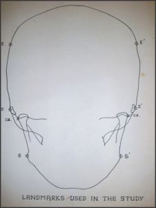
Cephalograms were traced on acetate paper and following landmarks were located:-
EURYON (E & E') : -A point located over each parietal bone at the greatest transverse diameter measured in the horizontal plane above the supero-mastoid and zygomatic crest (Salzmann 1966)[12]
ZYGION (Z & Z') : -The most lateral point on the zygomatic bone. (Sassouni 1957)[7]
Lateral Pole Of The Mandibular
CONDYLE (Cd& Cd') : The most lateral projection of the mandibular condyle.
GONION ( G & G') : The most lateral point at the gonial region in the horizontal plane (Richardson 1967)[13].
 |
 |
 |
|
 |
|
 |
|
The following widths were measured on the tracings obtained from the cephalograms:
1. INTERCONDYLAR DISTANCE (A) : Maximum transverse distance measured between laqteral poles of the two mandibular condyles (Cd- Cd').
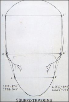
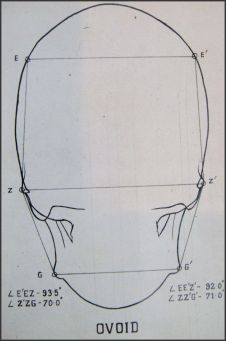
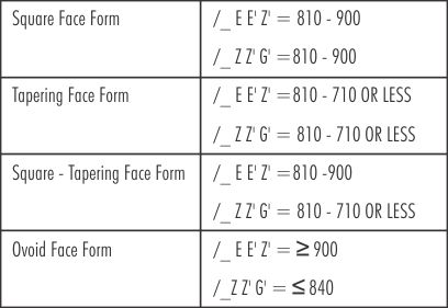
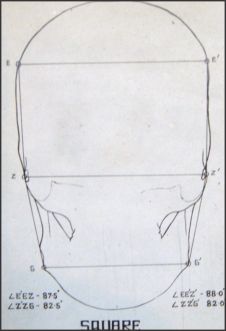
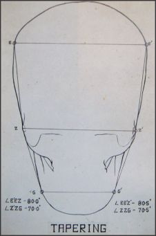
2. Determination Of Skeletal Face Form-
 |
|
 |
|
 |
|
Where:-
< E E' Z' - is the angle formed at the point Euryon by the lines joining the corresponding Zygion and Euryon on the other side ( upper face angle).
< Z Z' G'- is the angle formed at the point Zygion by the lines joining the corresponding Gonion and Zygion on the other side (lower face angle)
Observations:
1. It was observed that the ratio of "Bicondylar Width" (A), "Intermolar" (B) and "Intercanine" widths in overall sample was = 4.02 : 1.81 : 1.
2. For males this correlation ratio was= 4.02 : 1.79 : 1.
3. For females this ratio was = 4.02 : 1.83 : 1 .
4. There was a statistically significant difference (P = 0.001) between the mean values of three width measurements of males and females. Mean values of all the measurements were found greater for males.
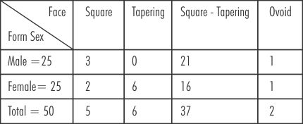
5. Distribution of subjects according to skeletal face form was as follows:-
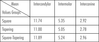
6. Three width measurements on the basis of skeletal face forms were as follows:-
*a very small sample of ovoid face form (2) was found, hence not considered significant for computing and tabulation
7. The ratio of above observations was:-
Discussion:
The present study was based upon the hypothesis that there exist varying correlations among various craniofacial and dental arch width dimensions; there could be a positive correlation between dental arch width and intercondylar distance.
This would provide an objective guideline to the Prosthodontist for the optimal placement of artificial posterior teeth particularly in mutilated and challenging cases.
The observation of overall samples when analyzed as a whole, the mean values of intercondylar, intermolar and Intercanine widths showed a ratio of 4.02: 1.81: 1. When same ratio was analysed separately for males and females, almost similar ratio was observed with a negligible variation of + 0.02 in intermolar widths.
The mean ratio for intermolar width in females (1.83) was higher as compared to males (1.79), was due to the fact that the mean values of all the three measurements were lesser than those in males; & this was because the Intercanine width in females was lesser in proportion, the effect of which on ratio showed apparent higher value of intermolar width. This signifies that the position of canines have direct bearing upon the dental arch form.
Furthermore the Intercanine and intercondylar distances showed constancy in proportions for overall samples as well as in gender variation. This signifies that the dental arch width in the canine region has more constant relationship with the intercondylar distance.
The mean values of three width dimensions were analysed in the three face forms. It was observed that the mean values of all the three measurements varied in different face forms. This showed the possibility of dominance of corresponding dental arch forms. The square and square-tapering groups showed significant positive correlation among the three dimensions.
Although the difference of mean values of square and square-tapering face forms was not significant, the tapering face group showed a significant difference which signifies the dominance of its typical arch form. These findings are in accordance to the findings of Agarwal, Tandon and Arora (1977)[16]. This shows that the ratios of the said dimensions is fairly constant for corresponding face forms.
A negative correlation for both sets of measurements was observed in tapering face group. Although it was found statistically insignificant, it could because of narrowing of arches with the increase in intercondylar distance. This seems to be an important finding of the study but needs to be further investigated.
A positive correlation was observed between intercondylar and intermolar width dimensions for the overall samples. This finding is in accordance to the findings of Frankel & Kronmann (1966)[11]. This confirms that intercondylar distance and dental arch width in 1st molar region have definite correlation between them.
A highly significant positive correlation was observed between intercondylar distance and Intercanine width for females. However an insignificant positive correlation was observed for the same in males. The cause of this variation need to be further investigated.
Conclusions:
To ascertain the possibilities of correlation between mandibular dental arch width and intercondylar distance, the present study was conducted on adult North Indian subjects. The following conclusions were drawn:-
1. A definitive correlation does exist between intercondylar distance and mandibular arch width.
2. The overall analysis showed more strong relationship of Intercanine width with intercondylar distance.
3. The three widths have a definitive ratio among them.
4. Different face forms have definite relationships for these measurements, typical among themselves.
5. In tapering face form, dental arch has a tendency to narrow down with increase of intercondylar distance and vice-versa.
References:
1. BONWILL W. G. A. (1889) - cited from " A consideration of normal arch form and some of the methods of determining it". Amer. J. Orthodontics, 5: 607- 729.
2. Williams P. N. (1917) - Determining the shape of normal arch. The Dental cosmos, 59: 695- 708.
3. Hardlicka A. (1940) - cited from "Normal variation of teeth, jaws and orthodonty", International journal of Orthodontics, 21: 1099- 1114.
4. Izard G. (1950)- cited from " Position of maxillary first permanent molar in the cephalofacial complex", Amer. J. Orthodontics, 43: 477- 509
5. Berger, H. (1929, 1952) - twenty-five years experience with zygomatic method, Amer. J. Orthodont. 38: 369-381.
6. Heneriques A. C. (1953)- the growth of the palate and the growth of the face during the period of changing dentition, Amer. J. Orthodont, 39: 836.
7. Sassouni V. (1955)- Roentgenographic cephalometric analysis of cephalo-facio-dental relationships. Amer. J. Orthodo. 41: 734-742.
8. Hixon E. H. , Meridith H. V. (1957)- An appraisal of two relationships. Amer. J. Orthodont. 43: 286-290.
9. Warren E. B. (1960) - A study of correlation of denture and skeletal widths, Amer. J. Orthonont. 46: 769-790.
10. Haspel L. S. (1962) - A study of arch width obtained from P-A cephalograms and plaster casts. Amer. J. Orthodont. 48: 394.
11. Frankel M. R. ; Kronmann J. H. (1966)- A cephalometric evaluation of craniofacial landmarks and their relationships to intermolar (mandibular) dimensions. Angle Orthodontist. 36: 263.
12. Salzmann J. A. (1966) Practice of Orthodontics.
13. Richardson M. E. (1967).The reproducibility of measurements on depressed posterior-anterior cephalometric radiographs. Angle Orthodontist 37: 48- 51.
14. Wei Stephen H. Y. (1970)- Craniofacial and dental widths. Angle Orthodontist. 40: 141.
15. Gupta D. S.; Dehadrai J. (1974) - Craniofacial and dental widths in north Indians. M.D.S. Thesis.
16. Agarwal N. K. ; Tandon B. K. ; Arora B.(1977)- A correlation between face-forms, arch forms and ridge forms. M. D. S. Thesis.
|
|
|
|
|
|
|