Introduction
Ossifying fibromas have formed an intriguing aspect of a variety of fibro-osseous lesions, probably owing to its wide histo-morphologic spectrum, including differentiation of bone, cementum and other calcifications and its differential diagnosis from fibrous dysplasia. It was Menzel in 1872, who first described this entity and in 1927 it was Montogmery who coined the term “Ossifying fibroma”. Ossifying fibromas occur in both central and peripheral locations of the jaw bones1. Histologically , the lesion is composed of varying amounts of immature and mature bony trabeculae, cementum like tissue, dystrophic calcifications, all in different configurations with varied stromal collagen content and cellularity. Collagen content of connective tissue has also been studied under polarized light using Picrosirius stain2. There appears to be no attempts to study the ossifying fibromas using van Gieson stain under polarized light, this study attempts to evaluate the stromal collagen with regard to fiber thickness & in various hard tissue elements of ossifying fibromas.
Aims and objectives of study
1) To evaluate the occurrence of various types of calcific deposits in ossifying fibroma.
2) To study the collagen fiber orientation, width, overall configuration in stroma and in calcific deposits ossifying fibroma.
Materials and methods
The present study was conducted in the Department of Oral Pathology, S.D.M Dental College and Hospital, Dharwad and included a histochemical study of 50 cases of Ossifying fibromas [25 cases central and 25 cases peripheral] inclusive of cemento-ossifying fibromas. Paraffin blocks of these cases were retrieved and non serial sections varying from 4 to 5 µm in thickness were cut on a semiautomatic Leica microtome and stained by Van Gieson stain. The corresponding hematoxylin and eosin stained slides were obtained for purpose of study and for comparison with Van Gieson stain. Staining results of van Gieson are as follows Nuclei-appear black colour, Collagen and bone- appear red colour, and other tissues including muscle and RBC'S-yellow colour. Both the Hematoxylin and eosin and van Gieson stained slides were then evaluated microscopically. Only Van Gieson stained slides, were viewed under polarized light microscopy.
Results
Table No. 1 and 2 explains the nature of stromal collagen in POF and COF cases respectively, as viewed under polarizing microscopy, using Van Gieson Stain. Thickness, colour, orientation and length of the collagen fibers were examined. Majority of the fibers observed in POF cases were a combination of thick and thin fibers (20/25 cases). Only 4 cases exhibited predominantly thin fibers and 1 case showed predominantly thick fibers. Color ranges from green to orange red. (Fig 1 and 2)
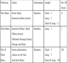 | TABLE NO.I Study of stromal collagen in POF under polarizing microscopy (van gieson stain)
 |
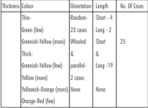 | TABLE NO.2 Study of stromal collagen in COF under polarizing microscopy (van gieson stain)
|
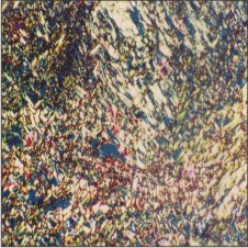
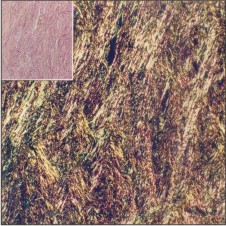 | FIGURE 1. Photomicrograph, showing thick, long, greenish yellow (few), and many yellowish orange and orange red stromal collagen fibers, in a whorled arrangement. (PL,VG, x 100) inset shows the same under lm (VG, x 100)
|
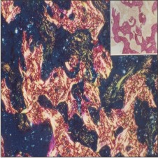 | FIGURE 2. Photomicrograph, showing thick, yellow and yellowish orange in a quilt pattern, in a curvilinear deposits. ( PL, VG, x100) inset shows the same in lm (VG, x 100)
|
Table No. 3 explains the total number of cases showing each type of hard tissue product elaborated and the osteoblastic and osteoclastic activity in both POF and COF cases. Both H & E and Van Gieson (VG) stained sections were examined and compared. Since there was no difference in the identification of the hard tissue elements between the H & E and VG stained sections, observations of both are considered under a single heading. The hard tissue elements were categorized as being osteoid, woven bone, lamellar bone, spheroid and curvoid deposits, curvilinear trabeculae based on the observations made by Eversole L R et al (1985)15.
![TABLE NO.3 Number of POF & COF cases showing each of the different hard tissue products and cell activity [h&e and van gieson (vg) stain]](article-image-1582-TABLE_NO.3_NUMBER_OF_POF_AND_COF_CASES_SHOWING_EAC.jpg) | TABLE NO.3 Number of POF & COF cases showing each of the different hard tissue products and cell activity [h&e and van gieson (vg) stain]
|
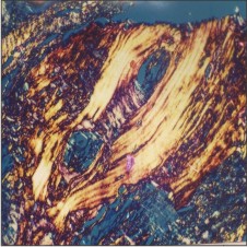
Table No. 4 shows the nature of collagen in various hard tissue elements in POF studied under polarization microscopy using Van Gieson stain. Osteoid showed thin and thick fibers, in random arrangement and were greenish yellow and yellow in colour. Woven bone, exhibited both thin and thick fibers in cross-hatched arrangement, with the thin fibers in the greenish yellow range. The majority of thick fibers were yellow coloured and few were greenish yellow. In lamellar bone, more thick fibers were seen in a parallel or lamellar arrangement and were yellow and orange red coloured. Very few thin yellow coloured fibers were noted. (Fig 3 and 4)
All the COF cases (Table No.5) showed both thin and thick fibers (25 cases) and majority of the cases showed both short and long fibers (19/25) and were in random arrangement in 23/25 cases. 2 cases, however exhibited a whorled pattern and parallel arrangement. The polarization colour of majority of the thin fibers was greenish yellow, with few in the green colour and the majority of the thick fibers showed yellow to yellowish orange and few with greenish yellow to orange red polarization colours. (Fig 2 and 5)
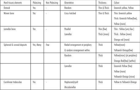 | TABLE NO.4 Study of collagen in hard tissue elements in peripheral ossifying fibromas under polarizing microscopy (van gieson stain)
|
 | FIGURE 3. Photomicrograph showing few thick, short, greenish yellow & orange red fibers & many thick, short & yellow fibers in random arrangement. Few thin, short, green to greenish yellow stromal collagen fibers in random arrangement are also seen. (PL,
|
 | FIGURE 4. Photomicrograph showing thick, long, yellow, yellowish orange and orange red stromal collagen fibers, in parallel arrangement. (PL, VG x 100). Inset shows the same under lm. (VG, x 100)
|
 | FIGURE 5. Photomicrograph, showing thick, yellow, broad lamellar pattern of collagen fibers, in lamellar trabecular bone. (PL, VG, x 250)
|
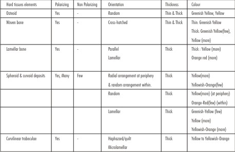 | TABLE NO.5 Study of Collagen In Hard Tissue Elements In Central Ossifying Fibromas Under Polarizing Microscopy (Van Gieson Stain)
|
Discussion
Ossifying fibromas have been queried repeatedly, to gain a better insight into the nature of the various hard tissue configurations. Though the various hard tissue products have been observed in light microscopy, attempts were made to study with the help of polarization microscopy3,4,5,6 to the birefringent nature of collagen and because it forms the frame work of all hard tissue elements. It was noted that based on collagen, cementum could be differentiated from bone6. All these studies were based on H & E sections. Since Van Gieson, stains collagen selectively, this study was undertaken to study the collagen content both in the stroma & in various hard tissue elements of ossifying fibromas (OF) in depth, under Light (LM) and Polarizing Microscopy (PM).
When VG sections were studied under polarizing microscopy, osteoid showed thin and thick fibers in random arrangement and were greenish - yellow and yellow in colour. Woven bone exhibited both thin and thick fibers in cross-hatched arrangement, with the thin fibers in the greenish-yellow range. The majority of thick fibers were yellow coloured and few were greenish yellow. In lamellar trabecular bone, more thick fibers were seen in a parallel or lamellar arrangement and were yellow and orange red coloured. Very few thin yellow coloured fibers were noted. Many of the spheroid and curvoid deposits polarized. 3 patterns were observed.
The first type, showed birefringence of peripheral collagen fibers and were in a radial arrangement (fringe), being yellow and yellowish orange coloured and composed of thick fibers. Very few thick fibers also polarized faintly within the masses, were in random arrangement and yellowish orange coloured.
In the second type, intersecting \ randomly arranged, short thick orange red fibers were seen within the mass, with more yellow coloured fibers at periphery. The third type showed lamellar orientation composed of thick fibers of yellow and yellowish orange colours & few of greenish yellow colours. Curvilinear trabeculae were made up of thick fibers in a quilt pattern or haphazard orientation and were thick yellow and yellowish orange.
On comparing POF & COF, lamellar bone showed only thick fibers more in the yellow and orange red range in COF cases. Otherwise, both POF and COF cases, exhibited same collagenous nature of hard tissue elements.
Polarization study of hard tissue elements have been evaluated so far, only in H&E sections and are observed as only dark & light lines of birefringence. Broad lamellar parallel fiber orientations (dark and light lines) were seen in lamellar bony trabeculae. A cross hatched fiber pattern 4, 3, 9 or a random pattern 6 with parallel oriented fibers but showing no lamellation was seen in woven bone. The dark and light lines of parallel birefringence in bony trabeculae were more widely spaced than those in cementoid tumor6. Spheroid, curvoid deposits (cemental deposits) seldom polarized and showed non specific birefringence with peripheral radiating fibers \ brush border \ brightly birefringent feather edge periphery 4, 6, 17 directed at right angles to the nidus. Fused globular cemental masses presented a mixed pattern of swirls and occasionally intersecting groups of short and thick fibers17. Many spheroid deposits, failed to polarize but some deposits revealed a radial fringe under polarized light resembling sharpey's fibers, and were similar to normal cementicles3. The appearances of calcifications were consistent with cementum.
The acellular basophilic type (type I) revealed a paucity of collagen and a brush border. The second type, showing less basophilia and with cellular inclusions but with no osteoblasts showed a quilted pattern and a parallel fiber pattern. The third type showing an eosinophilic dense cellular cemental mass having the appearance of bone, presented a quilt pattern, a coalescing globule pattern, or a parallel fiber pattern. In addition, these trabeculae were “molded” with rounded corners9. The curvilinear deposits exhibited a finely lamellar \ microlamellar \ haphazard \ quilted fiber pattern similar to cementum3, 4.Dystrophic calcifications showed a fairly diffuse birefringence with long, slender and parallel lines6.
Observation from our polarization microscopy study of hard tissue elements was similar to studies3, 4 except that of spheroid \ curvoid deposits. However the polarization colours could not be compared, as Van Gieson stain was not used in previous studies. Many of the spheroid \ curvoid deposits in our study on the contrary polarized and showed 3 different types of polarization. Our Type I collagen was in accordance with other studies3, 4 and the first type of cemental deposit described, and showing birefringence with peripheral radiating fibers9. In addition our study showed faint birefringence of few thick fibers within the masses, which were in random arrangement and yellowish orange coloured. This has not been observed by previous workers. Type II showed a prominent peripheral birefringence17, and type III both acellular & cellular9.
Based on picrosirius study of collagen19 and the collagen present in all the hard tissue elements were normal collagen, of type I and to some extent of type II20. Thus Van Gieson stain under polarizing microscopy, helps in delineating the nature of collagen apart from studying its orientation, fiber thickness and collagen thus stained, stands out clearly in hard tissue elements, compared to H & E sections under polarization microscopy. Thus polarizing microscopy helps in identifying hard tissue better than conventional microscopy9.
The lesions showing mainly spheroid / curvoid deposits and curvilinear trabeculae were considered to be cementoid lesions and lesions with an admixture of the above with osseous tissue as cemento-ossifying fibromas4. In accordance with it, in our study, Of 25 cases of POF, 7 cases were cementifying fibromas (CF), 3 were cemento-ossifying fibromas and the rest were OF and in 25 cases of COF studied, 4 cases were CF, 9 cases were cemento-ossifying fibromas and the rest were OF. But much against this myth, is the presence of ovoid calcifications in extragnathic lesions like meningiomas, prostatic adenocarcinomas etc 4. Curvilinear deposits representing cementum also have been seen in lesions other than cemental lesions like ossifying fibromas & fibrous dysplasias4.
A detailed study of fibro-osseous lesions of jaws in general showed that irrespective of them being reactive, or benignly neoplastic, they could involve factors which influenced differentiation along true osseous lines or by virtue of their anatomical proximity to periodontal structures were affected by factors which involved cemental differentiation9. The amorphous basophilic rounded calcified masses were considered to be cementum and though lesions containing such calcifications were designated as cementifying fibromas, it was difficult to explain the morphological difference between such cementum like masses and identical calcified masses seen in some fibro-osseous lesions of skull and in other bones. They concluded that though the lesions continue to be diagnosed on the basis of the dominant calcified part, there was no conclusive evidence as to whether the calcified masses in their cases were cementum or bony tissue but they diagnosed it to be a cemento ossifying fibroma based on previous observations21.
Though COF & CF to represent 2 facets of the same tumor, with one arising from osteoblasts & the other from cementoblasts8,22 differentiated cementum & bone based on their birefringent patterns under polarized light, it has not been possible to entirely separate these lesions. The findings of ovoid calcifications characteristic of cementum in any ossifying tumor of membrane of bone & not confined to the jaws 13, led to conclude that these calcified globules were not necessarily cementum and were not specific to lesions that originate in periodontal membrane and since the so called cemental globules could be seen in fibro-osseous lesions in all membrane bones; it was unrealistic to separate the ossifying & cementifying lesions.
Though, some studies clearly point out the differences between cementum and bone, both under light and polarizing microscopic observations3, 4, 6. Regarding spheroid, curvoid deposits in cementum recorded as nonspecific birefringence with only peripheral polarization 3.
Our observations & those of other studies showed birefringence within the mass in various patterns and some contained cellular inclusions. Even the basophilic masses showed occasional cell inclusions 9, 17. The analysis observed similar phenotypic expression - similar receptors, similar staining pattern with monoclonal antibody E11, of osteoblasts, osteocytes and cementoblasts & cementocytes, associated with cellular cementum. However the cells of primary cementum, showed a different phenotypic expression. Differences in their origins were also noted, with cellular cementum being formed by cells migrating from non-follicular sources like endosteal spaces. Hence, keeping observations of cementoid masses in extragnathic lesions and our and other author's findings of polarization microscopy of spheroid/curvoid deposits & curvilinear trabeculae, one strongly believes that cemental masses are nothing but bone tissues showing varied fiber content and orientations and sometimes containing cells23. Thus, there appears no point in exhausting our intellectual potency to classify OF into cementifying lesions & cemento-ossifying lesions and aptly consider all of them under the general heading of Ossifying fibromas of jaws.
Conclusion:
Polarization studies of stromal collagen in both POF and COF were essentially the same in that they showed the presence of normal collagen, belonging to mainly type I and III, with no pathologic types noted. Such an analysis appears to be unreported earlier.
Lamellar trabecular bone and woven bone in COF were seen more frequently than in previous studies. Spheroid, curvoid deposits occurring as single hard tissue product in COF was seen more than in previous studies. More of spheroid and curvoid deposits polarized in our study and showed 3 types of polarization patterns. The configuration of collagen in other hard tissues was similar to previous studies. Polarization study of hard tissue deposits, stained with Van Gieson appears to be unreported earlier.
References
1. Walter J M, Terry B C, Small E W,. "Aggressive ossifying fibroma of the maxilla": Review of the literature and report of case. Oral Surg1979, 37: 276-286.
2. Tomeo C, "Benign Cemento-ossifying fibroma of the mandible: A dilemma in Diagnosis". NYS Dental Journal 1978: 438-440.(46)
3. Eversole L R, Leider A S, and Nelson K, "Ossifying fibroma": A clinicopathologic study of sixty-four cases. Oral Surg Oral Med Oral Pathol1985, 60: 505-511.
4. Eversole L R, Sabes W R, and Rovin S, "Fibrous dysplasia: A nosologic problem in the diagnosis of fibro-osseous lesions of the jaws".J Oral Pathol 1972, 1: 189-220.
5. Giansanti J S, "The pattern and width of the collagen bundles in bone and cementum". Oral Surg1970, 30: 508-514.
6. Hamner J E, Scofiled H H and John C," Benign fibro- osseuos jaw lesions of periodontal origin".Cancer1968, 22: 861-878.
7. Neville B W, Damm D D, Allen C M, and Bouquot J E, "Oral & Maxillofacial Pathology 1995". W B Saunders Company.
8. Shafer, Hine and Levy, "A Text of Oral Pathology". 2nd Chapter, 'Benign and Malignant Tumours of the Oral Cavity'1993. W.B.Sounders Company, Philadelphia.
9. Waldron C A and Giansanti J S,. "Benign fibro-oseous lesions of the jaws: A clinical radiologic histologic review of sixty-five cases part II Benign fibro-osseous lesions of periodontal ligament origin" Oral Surg 1973, 35: 340-350.
10. Soames J V and Southams J C,. "Oral Pathology". Oxford Medical Publications 1998, New York.
11. Regezi J A and Sciubba J J, "Oral Pathology-Clinical Pathologic Correlations" 3rd edn 1999. W. B. Saunders Co. page-357-360.
12. Buchner A and Hansen L S, "The histomorphologic spectrum of peripheral ossifying fibroma". Oral Surg Oral Med Oral Pathol 1987, 63: 452-61.
13. Langdon J D, Rapidis A D and Patel M F, "Ossifying fibroma - one disease or six"? Analysis of 39-fibro-osseous lesions of the jaws. Br J Oral Surg 1976, 14: 1-11
14. Van Heerden WFP, Raubeen HEJ, Weir RG and Kredler J, "Giant ossifying fibroma": A clinico-pathologic study of 8 tumors. J Oral Pathol Med 1989, 18: 506-509.
15. Bradley E S and Leake D, "Ossifying fibroma involving maxilla and mandible". Report of case. Oral Surg Oral Med Oral Pathol 1968. 26: 605-614.
16. Carlisle J E and Hamner W B, "Giant Central ossifying fibroma of the mandible": Report of case .Oral Surg 1979, 37: 206-211.
17. Waldron C A, Giansanti J S and Browand B C, "Sclerotic central masses of the jaws (so-called chromic sclerosing osteomyelitis, sclerosing ostitis, multiple enostosis, and gingantiform cementoma)". Oral Surg 1975, 39: 590-604.
18. Amies A and Fleming W E, "Central ossifying fibroma of the jaws."Oral Surg Oral Med Oral Pathol 1962, 15: 1409-1414.
19. Dayan D, Hiss Y, Hirshberg A, Bubis J J and Wolman M,. "Are Polarization colors of picrosirius red stained collagen determined only by the diameter of the fibers? Histochemistry1989, 93: 27-29.
20. Junqueira L C U, Montes G S, and Sanchez E M,. "The influence of tissue section thickness on the study of collagen by the picrosirius polarization method". Histochemistry1982, 74: 153-156.
21. Takeda Y and Fyioka Y, "Multiple cemento - ossifying fibroma". Int J Oral Max Surg 1987, 16: 368-371.
22. Giansanti J S,. "The effects of acids upon the pathologic interpretation of the widths of the collagen bundles in bone". Oral Surg 1970, 30: 151-157.
23. Tencate A R, "Development of the Periodontium 1998" chapter no.12, Oral Histology Development, Structure, and Function. Mosby. |