Introduction
Maxillary anterior spacing or diastema is a common aesthetic complaint of patients and is frequently seen in children especially in the mixed dentition stage. Keene described midline diastema as anterior midline spacing greater than 0.5 mm between the proximal surfaces of adjacent teeth.1 He reported that the incidences of maxillary and mandibular midline diastema are 14.8% and 1.6%, respectively. Midline diastema may be considered normal for many children during the eruption of the permanent maxillary central incisors.
Taylor reported the incidence of midline diastema in 5 year olds as high as 97 per cent, and seen decreasing with age.2,3 Kaimenyi determined the prevalence of midline diastema and frenum attachments among school children (4-16 years) in Nairobi, Kenya. The commonest location of frenum attachment among children with lower midline diastema is at the mucogingival junction (86%), whereas with upper midline diastema to gingival region (50%). It was concluded that the maxilla had a higher prevalence of midline diastema than the mandible.4 Nainar and Gnanasundaram studied nearly 9774 patients in the age group of 13-35 years in South India (Chennai) and reported an incidence of true maxillary midline diastema (1.6%), which was greater than that of true mandibular midline diastemas (0.3%). 5
Angle concluded the presence of abnormal frenum as the cause for midline diastema and this view was supported by other researches.6, 7, 8, 9 Tait stated that the frenum is an effect and not a cause for the incidence of diastema and reported other causes such as ankylosed central incisor, flared or rotated central incisors, anodontia, macroglossia, dento-alveolar disproportion, localized spacing, closed bite, facial type, ethnic and familial characteristics, inter-premaxillary suture, and midline pathology.10 Weber listed the causes for spacing between the maxillary incisors as: a result of high frenum attachment; microdontia; macrognathia; supernumerary teeth; peg laterals; missing lateral incisors; midline cysts and habits such as thumb sucking, mouth-breathing and tongue-thrusting.
An accurate diagnosis is necessary before treatment can be initiated. No treatment should be initiated if the diastema is physiological and usually if the canines have not erupted. Different treatment modalities for midline diastema include removal of aetiology and simple removable appliances incorporating finger springs or split labial bow. Gleghorn reported a direct composite restoration technique to correct unaesthetic diastema.11 Follin ME reported extraction of mesiodens subsequently followed by the space closure utilizing simple fixed orthodontic therapy.13 Nakamura et al reported a ceramic restoration of anterior teeth without proximal reduction. 12
Symptoms
A diastema that occurs because of a mismatch between the teeth and the jaw does not have symptoms. However, spaces caused by a tongue thrust habit or periodontal disease will tend to expand or grow with time. The teeth may become loose, and discomfort or pain may occur, particularly during biting or chewing.
Prevention
Not all spaces can be prevented. For example, if the reason for a space is a missing tooth or a mismatch between the teeth and the jaw size, the spaces cannot be prevented without treatment.
Maintaining the gingival health is essential to good oral health. Regular flossing and brushing will help to prevent periodontal disease and its related bone loss.
People with a tongue thrust habit can re-learn to swallow by pushing their tongue up against their palate. Breaking this habit can prevent widening of the spaces between teeth.
Treatment Options:
Sometimes, a diastema is part of a set of problems that require orthodontic treatment. In other cases, a diastema is the only problem. However, some people may seek treatment for reasons of appearance.
Some people get braces, which move the teeth together - on both the upper and lower teeth. That's because moving any teeth affects the entire mouth.
If lateral incisors are too small, dentist may suggest widening them using crowns, veneers or bonding.
If there is a space because of missing teeth, it might need more extensive dental repair. This might include dental implants, a bridge or a partial denture.
If a large labial frenum is causing the gap, the frenum can be reduced through surgery called a frenectomy. If a frenectomy is done in a younger child, the space may close on its own. If it is done in an older child or an adult, the space may need to be closed with braces.
If the gap is caused by periodontal disease, then periodontal treatment by a dentist or gum specialist (periodontist) is necessary. When gum health is restored, in many cases braces can be used to move the teeth into place. A splint can be used to attach teeth to other teeth and prevent them from moving again. In some cases, a bridge will be required to close the spaces.
Case Reports:
Case 1
A 29-year-old female patient reported to the Department of Periodontics U.P. Dental College and Hospital, Lucknow; with a chief complain that she suffered for many years from shyness and self-consciousness as a result of the large diastema between her front teeth. (Figure 1).
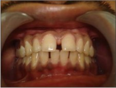 | Fig 01 - Pre-operative Diastema in 11,21.
 |
The patient's medical history did not reveal any systemic diseases. Intra-oral examination revealed presence of midline spacing between maxillary central incisors (4 mm).Patient was familiarized with the procedure to be followed and the expected outcome. Thorough oral prophylaxis was carried out, and crown reduction was done on the distal aspect of 11&21 for metal ceramic crowns. Rubber base impression was taken for both the upper and lower arc, and shade was noted (Figure 2). In the next appointment, metal ceramic crowns were cemented (Figure 3).
 | Fig 02 - Crown reduction -11,21on distal aspect.
|
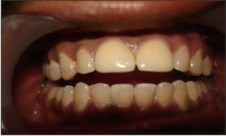 | Fig 03 - Post-operative Metal ceramic crown placed in11, 12.
|
Case2
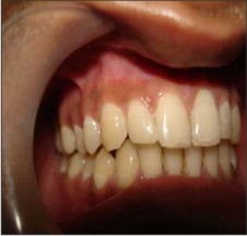
A 22-year-old female patient reported to the Department with a chief complain that she did not like the spaces between her teeth. Facially, the diastemas were not as apparent, but she was aware of the spaces and wanted the problem to be corrected (Figure 4).
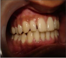 | Fig 04a - Pre-operative Diastema present between12-11&21-22.
|
 | Fig 04b - Pre-operative Diastema present between12-11&21-22.
|
She felt the appearance of her smile would improve if the space would be closed. During the consultation, treatment options were reviewed and the decision was made to correct the diastemas using a direct composite system (Direct Esthetic Composite System, IvoclarVivadent). A conservative approach was necessary, and emphasis was given to the health of the surrounding dentition and the patients overall satisfaction with the existing color and shape of her teeth.
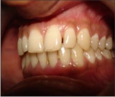
The tooth was isolated, acid etched, bonded and a composite resin buildup was performed to give the tooth the desired shape. Final finishing was done to establish the esthetic contour.
Case 3
A 19-year-old patient reported to the Department with the chief complaint of spacing in the upper front tooth region (Figure 5).
 | Fig 05a - Post-operative After Diastema closure by composite restoration.
|
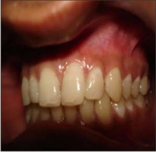 | Fig 05b - Post-operative After Diastema closure by composite restoration.
|
The patient's medical history did not reveal any systemic diseases. Intra-oral examination revealed presence of high frenal attachment and midline spacing between maxillary central incisors (4 mm).
A simple diagnostic test, i.e., blanching test was performed for an abnormal high frenum by observing the location of the alveolar attachment when intermittent pressure was exerted on the frenum (Figure 6).
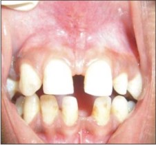 | Fig 06a - Midline Diastema in11, 12 Pre-operative High frenal attachment.
|
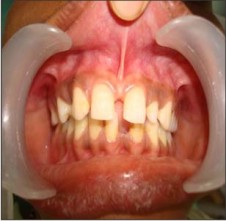 | Fig 06b - Midline Diastema in 11, 12 Pre-operative High frenal attachment.
|
A heavy band of tissue with a broad, fanlike base was attached to the palatine papillae that produced blanching of the papilla, which was safe to predict that the frenum would unfavorably influence the development of the anterior occlusion. After obtaining informed written consent from the parents, decision was made to remove high frenal attachment by a surgical technique.
Frenectomy was carried out under local anaesthesia with incision using No. 11 Bard Parker blade. In this technique, lateral incisions were made on either side of the frenum to the depth of the underlying bone. The free marginal tissues on the mesial side of the central incisors were not disturbed. The wedge of tissue was picked up with tissue forceps and excised with tissue shears at the area close enough to the origin of the frenum to provide a desirable cosmetic effect. Sutures were placed to identify the free tissue margins on either side of the removed tissue, and periodontal pack (Coe-pak) were placed for a week (Figure 7)
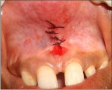 | Fig 07 - Sutures placed after surgery.
|
The patient was advised to return after a week for suture removal and periodical follow-up once a month, high frenal attachment and midline spacing for a period of 2 months, after which it was followed by orthodontic bracket placement.at the end a remarkable improvement in the aesthetics was observed, due to spontaneous closure of midline diastema (Figure 8).
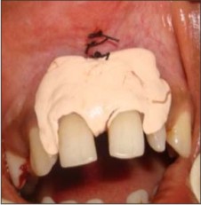 | Fig 08 - Periodontal pack (Coe-pak) placed.
|
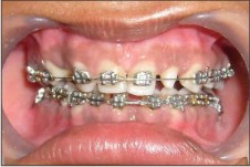 | Fig 09 - Orthodontic brackets placed.
|
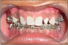 | Fig 10 - After Diastema closure in 11, 12.
|
Discussion
A diastema is a space or “gap,” most often seen between the two upper front teeth. At some stages of dental development, it is normal to have a diastema but it eventually closes during further development. There may be a number of reasons why patients present requesting diastema closure, including aesthetic and periodontal factors. With aesthetically compromised smiles, patients may become self-conscious, experience low self-esteem, and / or cover their mouths with their hands while speaking. Also, when diastemas result from significant tissue loss, which can change the airflow between the teeth, the reason for requesting diastema closure may be to correct obvious phonetic problems, particularly with s and th sounds.
Treatment of diastema varies and it requires correct diagnosis of its aetiology and early intervention relevant to the specific aetiology. Correct diagnosis includes medical and dental history; radiographical and clinical examinations and possibly tooth size evaluation. No treatment is usually initiated if the diastema is physiological / transient as it spontaneously closes after the eruption of permanent maxillary canines (11-12 years).
In third case, an attempt was made to remove the aetiology, even though the patient was only 19-year-old, considering the fact that there will be maximum active mesial movement of tooth during eruption. This resulted in the spontaneous closure of the midline diastema in 2 months. The patient was followed up for 4 months during which there was no change in the closed midline space but there developed a new space between the central and lateral incisors, which could be due to the eruption of permanent canine (Figure 3).
The patient has been followed up through regular recall for monitoring any changes in the anterior region.
Generally abnormal frenal attachment may require removal either before orthodontic treatment or at the end of active treatment. The advantage of excision prior to orthodontic treatment is the ease of surgical access. If the surgery is performed before the orthodontic procedure, the scar tissue might impede the closure of diastema but the noted advantages of excision after orthodontic tooth movement is the scar tissue formation which to helps maintain closure of diastema. . Follin ME stated that in spite of the success and excellent results, orthodontists have had a problem in correcting dental abnormalities, one particular area, which lends itself to relapse, is the diastema between the incisors.13 The surgical correction of a diastema has been successfully accomplished without orthodontic treatment in patients excepting a rapid correction.
Conclusion
Many anterior aesthetic cases present themselves during the course of daily practice routine. Selecting the proper treatment involves (1) the usual challenges of addressing the needs and expectations of the patient, (2) determining what degree of change is necessary to achieve the desired result, and (3) anticipating whether or not the patient will accept the treatment plan. Many aesthetic cases fall into the direct veneering category. The beauty of the direct veneering technique is that it provides the dentist with the freedom to be creative using a simple restorative technique and affords the patient a treatment that is completed in 1 day with beautiful, aesthetic results.
The concepts surrounding the diastema closure presented in this article are not overly complicated. Key to realizing a beautiful restoration for this purpose is the use of composites with superior handling, consistency, and color accuracy, and a restorative method that is very simple. Instead of trying to find an enamel shade, dentin shade, and incisal shade as in a conventional shaded layering technique using an anatomical approach enables the selection of a value of enamel and the matching of the underlying dentin layer, allowing the color to radiate from within the restoration just as it occurs in a natural tooth.
References
1. Koora K, Muthu MS et al Spontaneous closure of midline diastema following frenectomy. Journal of Indian Society of Pedodontics and Preventive Dentistry 2007: 25: 23-6 (s)
2. Nainar SMH and Gnanasundaram N. Incidence and etiology of midline diastema in a population in South India. The Angla Orthodontics 1989: 59: 277-282. (s)
3. Oji C and Obiechina AE. Diastema in Nigerian society . Odonto- Stomatologie Tropicale 1994: 17(68): 4-6. (s)
4. Lacy AM. Application of composite resin for single-appointment anterior and posterior diastema closure.Pract Periodontics Aesthet Dent. 1998;10(3):279-86.
5. Helvey GA. Closing diastemas and creating artificial gingiva with polymer ceramics. CompendContinEduc Dent. 2002;23(11):983-996.
6. Tallents RH. Artificial gingival replacements.Oral Health. 1983;73(2):37-40.
7. Keene HJ. Distribution of diastemas in the dentition of man. Am J PhysAnthropol 1963;21:437-41.
8. Angle EH. Treatment of malocclusion of the teeth. 7th ed. S.S. White Dental Manufacturing Co: Philadelphia; 1907. p. 103-4.
9. McCoy JD. Applied Orthodontia. 2nd ed. Lea and Febiger: Philadelphia; 1946. p. 72,96-7.
10. Stones HH. Oral and Dental diseases. 2nd ed. E and S Livingstone Ltd: Edinburgh; 1951. p. 19-21,211.
11. Gleghorn T. Direct composite technique for a smile makeover. Dent Today 1997;16:40,42,44.
12. Nakamura T, Ohyama T, Wakabayashi K. Ceramic restorations of anterior teeth without proximal reduction: A case report. Quintessence Int 2003;34:752-5.
13. Follin ME. Orthodontic movement of maxillary incisor into the midline: A case report. Swed Dent J 1985;9:9-13. |