Introduction
Restoration / replacement of lost teeth and surrounding structures pose difficulty in treatment planning when available bone and teeth present are inadequate for retention and support of a prosthesis. The fabrication of prosthesis for the partially edentulous arch encounters a special challenge where soft and hard tissue interferences, multiple paths of placement, tilted teeth and deranged occlusion complicate the treatment plan. Oral diseases like carcinomas and cysts require partial or complete removal of involved structures which further challenges the prosthesis design. Important factors like number of abutments, periodontal health and angulations of the abutments, length of edentulous span, condition of overlying mucosa, quantity and quality of bone in edentulous area determine the selection of prosthesis for such patients. Additionally, patient requirements, expectations and affordability of the treatment procedure can't be neglected. This case report presents one such dilemma encountered while treating a patient who was operated for odontogenic keratocyst and later rehabilitated with a very long span fixed partial denture which failed over a period of time.
Case Report
A 34 year female patient reported to the Department of Prosthodontics, Crown and Bridge & Implantology, People's College of Dental Sciences and Research Centre , Bhopal with a chief complaint of pain with 47 and loose prosthesis in mandibular arch. The tooth was endodontically treated 7 yrs back with a history of recurrent swellings. Her past dental history disclosed that she was operated upon her lower jaw for odontogenic keratocyst 9 yrs back. Post surgery her lost teeth had been replaced by a long span metal ceramic fixed partial denture using 47, 35, 36, 37 as abutments. Extraoral examination showed a concave profile and deep mentolabial sulcus, slight asymmetry of face.
The TMJ was asymptomatic. The vertical dimension of occlusion was maintained. Intraorally a very long span metal ceramic fixed partial denture was present replacing 31, 32, 33, 34, 41, 42, 43, 44, 45, 46. The ceramic veneer on 34, 35 was chipped off and an occlusal perforation was visible with the retainer on 47. A polyp had formed inside the retainer. The fixed partial denture was mobile on the right side. 47 was tender to percussion. On radiographic investigation root stump of 47 was seen with periapical rarefaction (Figure 1). 47 was difficult to salvage hence the patient was advised removal of the fixed partial denture. Since the patient was reluctant for complete removal, the fixed partial denture was sectioned at connector between 34 and 35. The sectioned movable part was removed and the retainer on 35 was finished and polished (Figure 2). The underlying mucosa in the edentulous area and the gingiva with respect to 47 was swollen and inflamed (Figure 3). 47 was extracted . Patient was adviced gum massage, chlorhexidine gargles and tissue rest.
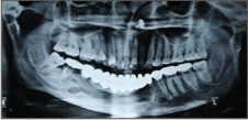 | Figure 1 : Orthopantomogram showing the long span fixed partial denture, resorbed alveolar bone in pontic area, secondary caries and periapical rarefaction with 47.
 |
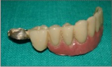 | Figure 2 : Sectioned part of the F.P.D.
|
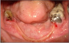 | Figure 3 : Intraoral condition after F.P.D.
|
A treatment prosthesis was prepared in the form of clasp retained acrylic resin partial denture (Figure 4). The Denture was relined with soft tissue conditioner ( Tissue Conditioner Kit, Medicept Dental, United kingdom) . 48 was mesially tilted and partially erupted so the abutment support and retention was inadequate. The shallow vestibular depth and insufficient supporting edentulous denture bearing area with bilateral lingual undercut compromised the retention of the prosthesis.
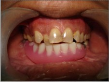 | Figure 4 : Treatment prosthesis in place, relined by soft tissue conditioner
|
The procedures for fabrication of flexible denture were performed after healing of the extraction wound and overlying mucosa in the edentulous area. The primary impressions were made in irreversible hydrocolloid material (Tropicalgin, Zhermack, Germany).
An individualized tray was prepared in auto polymerizing acrylic resin ( DPI- RR Cold Cure Acrylic Repair Material, Dental Products of India ,Mumbai , India) providing adequate relief for the thin crest of mandibular alveolar ridge. Border molding was performed using low fusing compound (DPI Pinnacle Tracing Sticks, Dental Products of India, Mumbai , India) .
Final impression of the mandibular arch was made using regular body (addition silicone) polyvinyl siloxane material ( Hydrorise, Zhermack SpA, 45021 Badia Polesine ( Rovigo), Italy) . Jaw relation was recorded and transferred to the articulator. Teeth selection and teeth arrangement were carried out. Patient's consent was obtained at the stage of try in. The denture was processed in flexible thermoplastic material (Valplast Int. Corp., US ) by injection molding. The denture was inserted and evaluated for comfort, fit, retention, stability , esthetics and speech (Figure 5 , Figure 6).
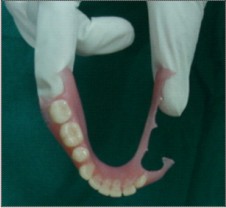 | Figure 5 : Valplast flexible denture
|
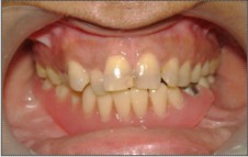 | Figure 6 : Valplast flexible denture placed in mouth.
|
Discussion
Odontogenic keratocyst is one of the most aggressive cysts of the oral cavity known for its rapid growth and high recurrence rate. The mentioned patient had undergone surgery for odontogenic keratocyst removal which resulted in deficiency of bone and teeth support required for the prosthetic replacement of lost structures.
While designing a prosthesis ascertainment of the availability of the tooth and bone support is imperative. Amongst many considerations Ante's law is given significance. Ante's law states that "the total periodontal surface of the abutment teeth must be equal to or larger than the surface of the replaced teeth".1 The law holds true if the periodontal condition of the abutment teeth is good. In the present case the Ante's law was overlooked which led to the ultimate failure of the prosthesis.2 As a result 47 had to be sacrificed further complicating the future treatment alternatives.
Treatment options available were preprosthetic ridge augmentation, implant supported prosthesis, cast partial denture, hybrid denture, flexible denture, conventional acrylic partial denture. The patient was apprehensive about any surgical intervention. The hybrid denture and cast partial denture would have required mouth preparation procedures to which the patient flatly refused. Patient had undergone innumerable dental procedures in the past which had made her skeptical and apprehensive.
In practice, sometimes patient's expectations and demands dictate the treatment plan. Treatment of choice remained was a flexible partial denture. The bilateral undercuts present in the mandibular arch required blockout of one of the undercuts or surgical intervention for easy placement and removal of the conventional removable partial denture. The blockout procedure cause the conventional denture flange to remain away from the alveolar ridge leading to food lodgement in that area. The flexible denture adapts itself well to the alveolar ridge avoiding the food entrapment.
The thermoplastic resins are used in dentistry for a broad variety of applications such as removable flexible partial dentures, preformed partial denture clasps, fiber reinforced fixed partial dentures temporary crowns and bridges, provisional crowns and bridges, obturators and speech therapy appliances, orthodontic retainers and brackets, impression tray 3 and border molding materials,4 occlusal splints, sleep apnea appliances, and implant abutments.
The flexible dentures can also be indicated in case of patients who has acrylic allergies, history of framework breakage , alternative to implants or fixed prostheses and presence of tori. There have been continuous efforts to improve the poly methyl methacrylate towards the enhancement of strength, better dimensional stability, better abrasion resistance and the achievement of radiopacity. 5, 6
Thermoplastic material for dental prostheses, Valplast (Valplast Int. Corp. - USA) , was first introduced to dentistry in the 1950s. The material is a Polyamide (nylon plastics).7,8 Valplast is nearly unbreakable, colored pink like the gums, can be built quite thin, and can form the denture base and the clasps as well. Since the clasps are built to curl around the necks of the teeth, they are practically indistinguishable from the gums that normally surround the teeth.
The resin is a biocompatible nylon thermoplastic with unique physical and aesthetic properties that provides unlimited design versatility and eliminates the concern about acrylic allergies. The flexible partial dentures permits the restoration to adapt to the constant movement and flexibility in the mouth. The flexibility, combined with strength and light weight, provides total comfort and esthetics. The flexible partial denture do not require mouth preparation procedures compared to conventional cast partial dentures.
The flip side of the valplast flexible dentures is the higher cost especially when the number of teeth to be replaced is more , compromised retention if remaining natural teeth are few, inability to reline .
There are some specific contra indications to flexible partial dentures. For example less than 4mm (all valplast) or 6mm (vitallium/valplast) of interocclusal space in the posterior area, bilateral free-end distal extensions with knife-edge ridges or lingual tori on the mandible, bilateral free-end distal extension on the maxilla with extreme atrophy of the alveolar ridges,d eep overbite cases (4mm or more) where the anterior teeth can be dislodged in excursive movements. 9
Conclusion
Muller De Van stated that "the preservation of that which remains is of utmost importance and not the meticulous replacement of that which has been lost".10 The dictum can't be understated. Proper selection of prostheses design is important for the long term success of the prostheses as well as the preservation and maintenance of the oral structures.
References
1. Ante IH. The fundamental principles of the abutments. Michigan Dental Society bulletin 1926 8;14
2. Leempoel PJB, Kayser AF, Van Rossum GMJM, De Haan AFJ . The survival rates of bridges. A study of 1674 bridges in 40 dutch general practices. J Oral Rehabil 1995;22: 327-330
3. Fujisawa M, Adachi K, Tsuruta S, et al. A procedure for fitting a fixed partial denture to an existing removable partial denture. J Prosthet Dent 2004;91(4):392-4.
4. Heath JR, Boru TK, Grant AA. The stability of temporary prosthetic base materials. J Oral Rehabil 1993;20(4):363-72.
5. Jagger DC, Harrison A, Jandt KD. The reinforcement of dentures. J Oral Rehabil. 1999;26:185-94.
6. Darbar UR, Hugget R, Harrison A. Denture fracture - A survey. Br Dent J 1994;176:342-5.
7. Lowe LG. Flexible denture flanges for patients exhibiting undercut tuberosities and reduced width of the buccal vestibule: a clinical report. J Prosthet Dent 2004; 92(2):128-31.
8. Phoenix RD, Mansueto MA, Ackerman NA, et al. Evaluation of mechanical and thermal properties of commonly used denture base resins. J Prosthodont 2004;13(1):17-27.
9. Michael Ditolla. Valplast, flexible esthetic partial denture, Chairside Clinical Perspective Clinical Techniques And Procedures April 2004 Volume 5, Issue 1. (Http://Www.Drditolla.Com/Pdfs/Valplast_Dentures.Pdf)
10. DeVan M. The nature of the partial denture foundation: suggestions for its preservation. T J Prosthet Dent. 1952;2(2):210-8. |