INTRODUCTION
Tooth transposition is defined as a unique and extreme form of ectopic eruption in which a permanent tooth develops and erupts in the position normally occupied by another permanent tooth.1Although the term ectopic eruption is often used in a wide sense to refer to any aberrant and abnormal eruption path taken by a tooth, the term transposition is confined to refer to an interchange in the position of two adjacent teeth within the same quadrant of the dental arch.2,3 It is a relatively rare developmental dental anomaly; the maxillary permanent canine is the tooth most frequently involved. Other dental anomalies, such as congenitally missing or peg-shaped maxillary lateral incisors, rotations and malpositions of the adjacent teeth, and retention of the deciduous canines, are most often associated with transposition.3,4,5,6 Transposition may be complete or incomplete.1,5,6 In complete transposition both crowns and entire root structures of the involved teeth are found in their transposed malposition. And in an incomplete transposition the crowns may be transposed, but the root apices still remain in their relative normal positions.1, 5, 6
Incidence and prevalence
Transposition may affect both sexes equally and, although it may occur in the maxilla or in the mandible, the frequency of maxillary permanent canine involvement is the greatest.1 In the maxilla the canine is transposed most frequently with the first premolar, less often with the lateral incisor followed rarely by central incisor or second premolar.1, 4 In the mandible transposition is reported to involve the canine and lateral incisor only.
It has been reported that transposition of maxillary teeth occurs approximately in one of 300 hundred orthodontic patients and that transposition between the canine and first premolar appears most often (70%) in maxillary dentition, followed by one between canine and lateral incisor (20%).2 ,3
Transposition has never been reported in both arches simultaneously and it has never been reported to occur in the deciduous dentition. Unilateral transpositions are found more often than bilateral transpositions and show left side dominence.2, 5
ETIOLOGY
Although there are several theories put forward, the etiology of transposition is still unclear. A possible explanation for tooth transposition would be an exchange in position between developing tooth buds and also genetic or hereditary factor can play a role. 3, 6 The maxillary canine is more likely than any other tooth to become impacted or transposed. The maxillary permanent canines has longest period of development and long way to travel from early formation stage to its complete eruption.7, 8 During its long eruption pathway, the canines move labially and mesially and can be palpated high in the labial sulcus.7 Observing the high incidence of maxillary canine transposition one possible cause may be prolonged retention of deciduous canines. Other causes of transposition include trauma, mechanical interference, bone disease, tumors or cysts.1, 7, 8
Following a multifactorial hereditary model, Peck et al2 and Ely et al10 suggested that transposition of a maxillary canine and first premolar is genetically controlled and also environment plays a role.2, 10 This conclusion was reached because of the moderate rate of bilateral occurrence, increased prevalence of additional dental anomalies as hypodontia, occurrence following a hereditary pattern, and varying prevalence among populations.2, 10
CLASSIFICATION OF TRANSPOSITION
Peck and Peck11 conducted a wide review of case reports of tooth transpositions in the maxillary arch and established a classification based on anatomical factors. From 201 case reports reviewed, the authors systematically classified transposition, in decreasing
order of frequency
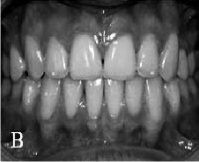 | Fig 1:Normal Dentition
 |
 | Fig 2:Transposition Of Canines To Premolar Region
|
 | Fig 3:Transposition Of Canines To Lateral Incisor Region
|
 | Fig 4:Transposition Of Canine To First Molar Region
|
 | Fig 5:Transposition Of Lateral Incisor To Central Incisor
|
 | Fig 6:Transposition Of Canine To Central Incisor Region
|
(Fig 2-6 depicts the possible transpositions that may usually occur,
Fig 1 shows normal Dentition):
1. Canine—first premolar (Mx.C.P1)
2. Canine—lateral incisor (Mx.C.I2)
3. Canine to first molar site (Mx.C to M1)
4. Lateral incisor—central incisor (Mx.I2.I1)
5. Canine to central incisor site (Mx.C to I1)
In the classification aberrant positioning of the maxillary canine is a feature of four of the five types, the exception being the special situation of lateral incisor-central incisor transposition.
TREATMENT CONSIDERATION
Early diagnosis of a developing transposition is extremely important and has a great influence on prognosis. This may usually be performed by a radiographic examination when the patient is between 6 and 8 years of age. When the alteration is detected early, interceptive procedures including extraction of deciduous teeth and placement of eruption guides for the permanent teeth may be performed, thus preventing complete development of the anomaly. On the other hand, when transposition is detected at a later stage, orthodontic treatment planning and intervention must be addressed.1, 3 Upper canine-premolar transposition in adult patients allows consideration of several treatment options, with or without extraction of the premolar.3, 12
In nonextraction treatment, generally the position of transposed teeth is maintained without restoring their natural tooth position. However, the upper canine-premolar transposed order provides esthetic and functional considerations. The differences in the size, shape, and tooth color between canine and premolar sometimes cause anterior esthetic problems. The gingival contour of the premolar is lower relative to the canine, requiring a periodontal gingival recontouring procedure. The palatal cusp of the transposed premolar might cause functional interference. The size and shape of premolar are completely recontoured to resemble a canine after root canal treatment .1, 12
Treatment with premolar extraction is considered as one of the alternatives. The transposed tooth is then moved to its natural position. This treatment approach is preferred when there is a severe arch length deficiency. Root interference needs to be considered. Root interference during tooth movement to correct tooth order tends to occur more frequently in canine-premolar transposition than in lateral-canine transposition. This probably occurs because the labiolingual width of a premolar is much wider than that of the lateral incisor.
In addition, gingival recession around the repositioned canines should be considered because of the long journey of canine through the dense buccal compact bone.1, 12, 13 The other problem in restoring the natural tooth order is prolonged orthodontic treatment due to difficulties in root movement.13 As a general rule, it is not advisable to correct a transposed tooth order because of insufficient buccopalatal width of bone support when two adjacent teeth are moving in different directions.1, 13
Summary
Tooth transposition is defined as the positional interchange of two adjacent teeth within the same quadrant. Tooth transposition is a rare positional anomaly that causes problems in both esthetic and functional aspect. Although the etiology of tooth transposition remains unclear, two principal theories of this anomaly have been proposed. One is migration of the tooth buds from the normal path during odontogenesis. The other is a genetic influence. The maxillary permanent canine is the tooth most frequently involved. Therefore, the maxillary canines should be regularly monitored after early loss of deciduous teeth. Transposition is also associated with other dental anomalies, such as congenitally missing or peg-shaped maxillary lateral incisors, rotations and malpositions of the adjacent teeth, and retention of the deciduous canines. Transposition may affect both sexes equally and shows higher maxillary prevalence. It is not seen in both the arches simultaneously and also is not seen in deciduous dentition. Unilateral transpositions are found more often than bilateral transpositions and show left side dominence. Treatment options for transposition include early diagnosis and guiding eruption to orthodontic management either by non-extraction or extraction modalities. When treating transpositions, especially MxC.P1, many factors that affect the treatment results must be considered, such as esthetics, occlusion, treatment period, patient comfort, patient cooperation, and periodontal support. For the achievement of optimal function and esthetics in cases with tooth transposition requires the utmost care in the design of the treatment.
References:
1. Shapira Y and Kuftinec MM. Maxillary canine-lateral incisor transposition. Am J Ortho. 1989 ; 95 : 5439 – 444
2. Peck L, Peck S, Atia Y. Maxillary canine - first premolar transposition, associated dental anomalies and genetic basis. Angle Ortho. 1993; 2: 99 – 110.
3. Filhoa LC, Cardosob MA, Anc TL and Bertozd FA. Maxillary Canine—First Premolar Transposition. Restoring Normal Tooth Order with Segmented Mechanics. Angle Orthod.2007; 77(1):167 – 175.
4. Shapira Y and Kuftinec MM. Orthodontic management of mandibular canine-incisor transposition. Am J ortho. 1983; 83 (4): 271 – 276.
5. Yýlmaz HH, Tu¨rkkahraman H and O¨ Sayýn M. Prevalence of tooth transpositions and associated dental anomalies in a Turkish population. Dentomaxillofacial Radiology. 2005; 34: 32–35.
6. Sato K, Yokozeki M, Takagi T, Moriyama K. An Orthodontic Case of Transposition of the Upper Right Canine and First Premolar. Angle Orthod 2002; 72(3):275–278.
7. Shapira Y, Kuftinec MM. Tooth transposition- review of literature and treatment consideration. Angle Ortho. 1989; 59(4): 271 -76.
8. Göyenç Y, karaman AI, Gökalp A. Unusual Ectopic Eruption of Maxillary Canines. J Clin Orthod. 1995 Sep; 29(9):580-2.
9. Tu¨rkkahraman H, zgu¨ r Sayýn, H. Yýlmaz HH. Maxillary Canine Transposition to Incisor Site: A Rare Condition. Angle Orthod. 2005; 75: 284 – 287.
10. Ely NJ , Sherriff M and Cobourne MT. Dental transposition as a disorder of genetic origin. Eur Jour Orthod. 2006; 28: 145–151.
11. Peck S and Peck L. Classifications of maxillary tooth transpositions. . Am J Ortho.1995; 107: 505 – 517.
12. Kurodaa S, Kurodab Y. Nonextraction Treatment of Upper Canine–Premolar Transposition in an Adult Patient. Angle Orthod 2005; 75:472–477.
13.Babacana H, Kilic B, Bicakci A. Maxillary Canine-First Premolar Transposition in the Permanent Dentition. Angle Orthod. 2008. 78(5): 954 – 960.
|