Introduction
A provisional restoration is a temporary dental restoration designed to enhance aesthetics and function for a limited period of time which is to be replaced by a definitive dental restoration1. Fabrication of provisional restorations is an important step toward achieving a successful fixed prosthodontic treatment.1, 2
Provisional restorations are needed to maintain gingival health and protect the pulp of prepared teeth.3-6. The term provisional may suggest that this step requires less stringent adherence to prosthodontic principles than other steps of crown or fixed partial dentures fabrication. However lack of attention at any step in the restorative procedure can yield disastrous results. An improper provisional restoration may result in the eventual loss of more time than that was initially thought saved. It is therefore essential that biological, mechanical and aesthetic requirements are satisfied by provisional restoration. The mean duration of a provisional restoration is seven to ten days, but sometimes clinical delay, disease, financial constrains etc may prolong the period for which it is in use. Therefore the success of fixed prosthodontics to a large extent depends on the care and method in which the provisional restoration is designed and fabricated.
Ideal requirements of provisional restorative materials are adequate working time, ease of mix and repair, bio compatibility with pulp and soft tissues, dimensional stability during and after fabrication, marginal adaptation,shade and color stability etc.
Out of these ,one of the important requirement of the provisional restoration is good marginal adaptation. Poor marginal fit allows the passage of fluids and bacteria into the gap and may predispose the tooth to caries or pulpitis. Therefore the objective of this in-vitro study was to compare the marginal accuracy of four provisional restorative materials commonly used in clinical practice.
Material And Methods
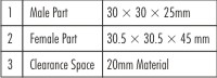
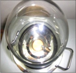
The present study was undertaken at department of Prosthodontics, Himachal Dental College Sundernagar. An in-vitro method was used to simulate a clinical technique in which the interim crowns were made directly on the prepared metal die using a matrix. For holding of this matrix and metal die this metal jig was fabricated as per dimensions given by Farhanz el al.8(Fig.1)
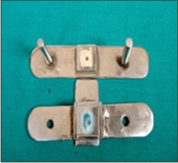 | Fig. 1 : Male And Female Parts Of Metal Jig
 |
Dimensional details of the apparatus:
Volumetric Dimension:
The provisional restorative materials used in the study are shown in Table A .
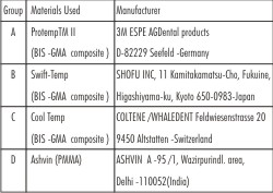 | Table A Provisional restorative materials and their manufacturers
|
An artificial maxillary central incisor (FRASECO German) was prepared to receive a complete crown restoration with a shoulder finish line. Putty reline technique was used to make the impression of the prepared tooth using elastomeric impression material. Inlay casting wax (Bego) was flown into the impression of that tooth. Root portion was built rectangular with the inlay casting wax. Then the wax pattern was retrieved from this impression along with the root portion. It was then invested and casted. The model retrieved after casting was finished and polished A matrix was prepared to hold the provisional restorative material over the prepared tooth. A crown analogue was prepared over the metal die used in the study.. The metal die with the crown analogue was mounted in the plaster of paris in the male part of the metal jig and polivinylsiloxane putty impression of the die was taken along with crown analogue. For this, an equal proportion of base and catalyst pastes of polivinylsiloxane putty impression material was mixed and placed in the female part of the jig. The female part was inverted over the male part till the mark of location on both parts coincided with each other .The polymerized impression served as the matrix for making the provisional restorations.The matrix was filled with various provisional crown materials used in the study respectively and was seated over the metal die to fabricate theprovisional crowns Fig 2.The gingival margins of all provisional restorations were trimmed with rubber wheel under a microscope at 10X magnification. Trimming of all interim restoration margins was performed by single operator. Then the crowns were made conductive by placing them in ion sputter coater Fig 3,4. Each provisional restoration along with the metal die was held in place with the help of a stub in the chamber of the scanning electron microscope Fig 5. The marginal discrepancy of each provisional restoration was determined by measuring the vertical space between the margin of the provisional restoration and the finish line of the metal die under Scanning Electron Microscope.
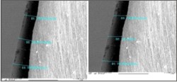
For each interim restoration, measurements were made on all four sides i.e. buccal, mesial distalandlingual, Fig 6, 7, 8, 9. On one given side a reference point at the middle of margin was marked. Three measurements were made on the either side of the reference point. Hence marginal gap on each surface was recorded at six different points and a mean of these six readings was obtained on each side. The data thus obtained was subjected to statistical analysis.
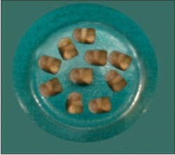
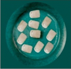 | Fig. 2 : Fabricated Samples
|
 | Fig. 3 : Loaded Sample In Sputter Coater
|
 | Fig. 4 : Samples After Gold Coating
|
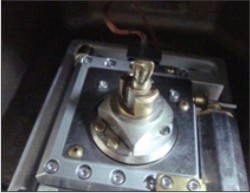 | Fig. 5 : Loaded Sample In Sem
|
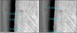 | Fig. 6 : Marginal Gap On Either Side Of The Reference Line On The Buccal Surface.
|
 | Fig. 7 : Marginal Gap On Either Side Of The Reference Line On The Mesial Surface.
|
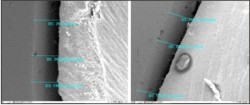 | Fig. 8 : Marginal Gap On Either Side Of The Reference Line On The Distal Surface.
|
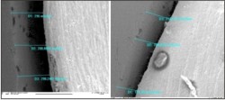 | Fig. 9 : Marginal Gap On Either Side Of The Reference Line On The Lingual Surface.
|
Observations
A total number of forty samples, prepared from four different kinds of provisional restorative materials, were evaluated for the marginal discrepancies. The data obtained was analyzed under following parameters :
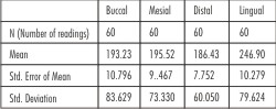
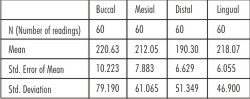 | Table 1 Vertical Mean marginal gap in (microns) of ProtemTM II (Group A)
|
Table 1 shows the mean marginal gap on each of the four surfaces of the provisional restoration obtained fromProtempTMII. The mean marginal gap is maximum on the buccal side i.e. (220.63) and minimum in distal (190.30).
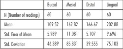
 | Table 2 Vertical Mean marginal gap in (microns) of Swift-Temp (Group B)
|
Table 2 shows the mean marginal gap of Swift-Temp on each of the four surfaces of the provisional restoration. The mean marginal gap is maximum on the lingual side i.e. (246.90) and minimum on distal (186.43).
 | Table 3 Vertical Mean marginal gap in (microns) in Cool Temp(Group C)
|
Table 3 shows the mean marginal gap fabricated with Cooltemp on each of the four surfaces of the provisional restoration .The mean marginal gap is maximum on the lingual side i.e. (202.88) and minimum in buccal (109.52).
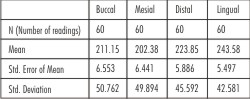 | Table 4 Vertical Mean marginal gap in (microns) in Ashvin(Group D)
|
Table 4 shows the mean marginal gap fabricated with Ashvin on each of the four surfaces of the provisional restoration. The mean marginal gap is maximum on the lingual side i.e. (243.58) and minimum in mesial (202.38).
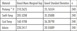 | Table 5 Grand Mean Marginal Gap and Grand Standard Deviation (microns) of the four provisional restorative materials
|
Table 5 represents the descriptive statistics .This table basically describes the Grand Mean Marginal Gap (microns) and Grand Standard Deviation of the four provisional restorative materials which were used in the study. Maximum Grand mean marginal gap is observed for Ashvin (220.2417) followed by :ProtempTM II 210.2625 ,Swift Temp 205.5208 and Cool Temp 160.4708
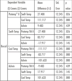 | Table 6 Pair wise Comparison (Buccal Surface)
|
Table 6 represents the pair wise comparison of the four different materials used in the study on the Buccal surface. The above stated table represents the mean difference, Std. error and significant difference. According to the above stated table,ProtempTM II shows the significant difference with Swift-Temp and CoolTemp i.e. (27.400 and 111.117) at 5% and 1% level respectively.
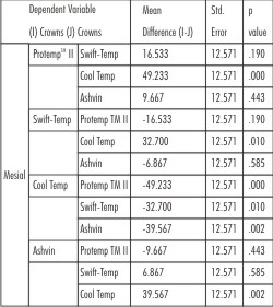 | Table 7 Pair wise Comparison (Mesial Surface)
|
Table 7 represents the pair wise comparison between the four different materials used in the study on the mesial surface. According to the above stated table the protempTM II shows the significant difference with Cool Temp i.e. (49.233) at 1% level. Similarly, it is also found that there is a significant difference between CoolTemp with Swift -Temp i.e.(32.700) and Ashvin(39.567) at 1% level.
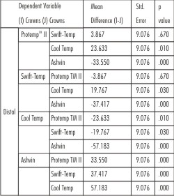 | Table 8 Pair wise Comparison (Distal Surface)
|
Table 8 represents the pair wise comparison between the four different materials used in the study on the distal surface. On the distal surface ProtempTMII, Swift-Temp and Ashvin shows a significant difference of (23.633, 19.767 and 57.183) respectively with the cool temp .
 | Table 9 Pair wise Comparison (Lingual Surface)
|
Table 9 represents the pair wise comparison between the four different materials used in the study on the lingual surface. There is an insignificant difference of (15.183) at 5% level between ProtempTMII and Cool Temp. As far as Ashvin and Swift Temp are concerned there is a significant difference of (40.700 and 44.017) at 1%level with Cool Temp. ProtempTMII shows significant difference with Swift-Temp ( 28.833)at 1% level and Ashvin (25.517) at 5%level.
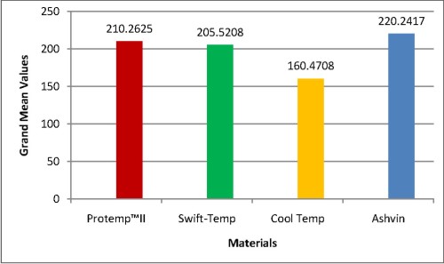 | Graph 1 : Grand Mean Marginal Gap
|
Discussion
Fabrication of provisional restoration is an important step towards achieving successful prosthodontic treatment. Provisional crowns are seated on the prepared abutments at a critical phase of treatmentthat is when gingiva is injured during crown preparation and any discrepancies in marginal adaptation of provisional restoration further complicates the situation. Therefore the most important morphologic and physiologic requirements for a provisional crown is suitable marginal adaptation. The purpose of this in-vitro study was to compare vertical marginal discrepancy of provisional crowns made from four provisional restorative materials. The four materials included in the study were different brands of chemically cured Bis-GMA resins and one PMMA resin.
On measuring the marginal discrepancy as seen in Table No 1-4 provisional restorations made under group D i.e. restoration made from Ashvin demonstrate significant increase in marginal gap size as compared to the other three materials tested on all the four surfaces .It is assumed that the observed marginal discrepancies were caused by the polymerization shrinkage of self cure resins. The results of this study were similar to the findings of study conducted by Robinson and Hovijitra4 who have attributed the marginal discrepancy to polymerization shrinkage. Similar results were obtained by Braden and Clark as well. 33
Table 5 describes the grand mean marginal gap and grand standard deviation of the four provisional materials used in the study. This can be seen that the grand mean marginal gap of cool temp is minimum as compared to other materials used in the study. It is further observed that the grand standard deviation of cool temp is minimum and maximum is of Ashvin. It depicts that Cooltemp material has the best marginal fit on all the surfaces.
The above mentioned facts are further clarified in Table no 6 -9 where pair wise comparison of different materials was carried out on all the four surfaces.From the comparisons done in Table no. 6-9 it was concluded thatCooltemp shows the best marginal fit on all the four surfaces followed byswift-temp protempand ashvin.
From the above mentioned observations it is clear that the provisional restorations made from Bis-GMA composite resins showed more close adaptation as compared those made with PMMA. This is due the fact that polymerization shrinkage is less in composite resins as compared to self cure resins .Moreover there is lack of exothermic reaction in composite resins which contributes to better adaptability .These results are comparable to previous studies conducted by Young et al30 and Farahanz et al8 where they have concluded that composite resins show less marginal discrepancy as compared to self cure resins.
Further it is clear from the observations that among the different composite resins used in the study Cool Temp showed least marginal discrepancy followed by Swift Temp and ProtempTMII.
This finding can be explained by the fact that Cooltemp was supplied as a cartridge based dispensing system with auto mix technique resulting in a more accurately proportioned and consistent mix.
In case of Swift Temp a calibrated mixing pad was provided on which equal lengths of base and catalyst pastes could be dispensed for a more accurate mix. Whereas in case of ProtempTM II base and catalyst were provided in separate cartridges and there were chances of error while dispensing the two pastes resulting into less accurately proportioned and consistent mix. This fact was emphasized by Bausch et al, in their study on polymerization shrinkage of composite resins where they concluded that accurate proportioning of base and catalyst pastes resulted intopolymerization shrinkage. Same results were obtained by Farahanz et al,8 where they analyzed that cartridge based dispensing system with auto mix technique resulted into less marginal discrepancy. So it is clear that the composite resin based provisional restorative materials show less marginal discrepancy as compared to self cure resins. However, in a study conducted by Barghi and Simmons on marginal integrity of the self cure resin temporary crowns it was concluded that if self cure resin has to be used as a provisional restorative material, a reline technique should be employed to improve the marginal integrity.
Summary and Conclusion
The primary objective of a provisional restoration is to maintain good gingival health prior to the placement of the final restoration. An accurate marginal fit of temporary crowns is an important factor that needs consideration as it protects the prepared teeth from physical, chemical, bacterial and thermal injury. Thus, the present study aims at studying the marginal accuracy of the provisional restorations and evaluating the best possible marginal adaptation amongst the commonly used materials.
It may be concluded from the present study that:
1) Bis-GMA provisional restorative material shows the better marginal fit and out of four materials used in studyCooltemp showed the best fit on all the surfaces
2) Ashvin (PMMA), on the other hand, showed a poor overall marginal fit as compared to other Bis-GMA based materials.
3) It can also be concluded that more accurately proportioned and consistent mix resulted in better marginal fit .
Refrences
1. Kim SH, Watts DC. Exotherm behavior of the polymer based provisional crown and fixed partial denture materials. Dent Mater 2004;20:383-7.
2. Haselton DR, Diaz Arnold AM, Vargas MA. Flexural strength of provisional crown and fixed partial denture resins. J Prosthet Dent 2002;87:225-8.
3. Barghi N, Simmons EW Jr. The Marginal Integrity of the temporary acryclic resin crown, J Prosthet Dent 1976; 36:274-7.
4. Robinson FB, Hovijitra S. Marginal fit of Direct temporary crowns. J Prosthet Dent 1982;47:390-2.
5. Rosenstiel SF, Land MF, Fujimoto J. Comtemporary Fixed Prosthodontics. 3rd ed. St. Louis: Mosby; 2000. p.391-4.
6. Federick DR. The provisional fixed partial denture. J Prosthet Dent 1975;34:520-6.
7. Dawson PE. Evaluation, Diagnosis, and Treatment of Occlusal Problems. 2nd ed. St Louis: Mosby; 1989. p. 288-9.
8. Nejatidaesh F, Lotfi R H, Ornid S. Marginal accuracy of interim restoration fabricated from four interim autopolymerizing resins. J Prosthet Dent 2006;95:364-7.
9. Tjan AH, Tjan AH, Grant BE. Marginal accuracy of temporary composite crowns. J Prosthet Dent 1987;58:417-21.
10. Tjan AH, Castelnuovo J, Shiostu G. Marginal fidelity of crowns fabricated from six proprietary provisional materials. J Prosthet Dent 1997;77:482-85.
11. Donaldson D. The etiology of gingival recession associated with temporary crowns. J Periodont 1974;45:468-71.
12. Micheal I, Mac Entee. A histological evaluation of tissue response to three currently used temporary acrylic crowns. J Prosthet Dent 1978;39:42-46.
13. Crispin BJ, Watson JF. Esthetic considerations for the placement of anterior crown margins. J Prosthet Dent 1980;44:290.
14. Gavilis JR, Morency JD. The effects of various finish line preparations on the marginal seal and occlusal seat of full crown preparation. J Prosthet Dent 1981;45:138-45.
15. Monday J.J.L, Blais D. Marginal adaptation of provisional acrylic resin crown. J Prosthet Dent 1985;54;194-97
16. Anthony HL Tjan ,Dr.Dent, Sarkissian R. Effect of preparation finish on retention and fit of complete crowns. J Prosthet Dent 1986;56:283-88.
17. Koumjian JH, Holmes JB. Marginal accuracy of provisional restorative material. J Prosthet Dent 1990;63:639-42.
18. Driscoll FC, Woolsey G, Ferguson MW. Comparison of exothermic release during polymerization of four materials use to fabricate interim restoration. J Prosthet Dent 1991;65:504-06.
19. Hung CM, Weiner S. Effects of thermocycling and occlusal forces on the margins of provisional acrylic resin crowns. J Prosthet Dent 1993;69:573-77.
20. Kern M, Schaller HG, Strub JR. Marginal fit of restoration before and after cementation. in vivo. Int. J. Prosthodont 1993;6:585-91.
21. Brent Moulding M, Robert W Loney. Marginal accuracy ofprovisional restoration fabricated by different techniques. Int J Prosthodont 1994;7:468-72.
22. Kern M, Schaller HG, Strub JR. Marginal fit of restoration before and after cementation in vivo. Int. J. Prosthodont 1993;6:585-91.
23. Zwetchkenbaum S, Weiner S. Effects of relining on longterm marginal stability of provisional crowns. J Prosthet Dent 1995;73:525-29.
24. Castelnuovo J, Tjan AHL. Temperature rise in pupal chamber during fabrication of provisional resinous crowns. J Prosthet Dent 1997;78:441-46.
25. Baldissara P, Comin G, Martone F, Scotti R. Comparative study of the marginal microleakage of six cements in fixed provisional crowns. J Prosthet Dent 1998;80:417-22.
26. Dubois RJ, Kyrialcatis P, Weiner S, Vaidyanathan TK. Effects of occlusal loading and thermocycling on the marginal gaps of light polymerized and auto polymerized resin provisional crown. J Prosthet Dent 1999;82:161-68.
27. Ogawa T, Aizqwa S, Tanaka M, Matsuya S, Hasegawa A, Koyana K. Effect of water temperature on the fit of provisional crown margins during polymerization. J Prosthet Dent 1999;82:658-66.
28. Beschnidt SM, Strub JR. Evaluation of marginal accuracy of different all- ceramic crown systems after simulation in artificial mouth. J Oral Rehabil 1999;26:582-93.
29. Ehrenberg DS, Weiner S. Change in marginal gap size of provisional resin crown after occlusal loading and thermocycling. J Prosthet Dent 2000;84:139-148.
30. Young HM, Smith CT, Mortan D. Comparative in vitro evaluation of two provisional restorative materials. J Prosthet Dent 2001;85:129-32.
31. Piemjai M. Effects of seating force, margin design and cement on marginal seal and retention of complete metal crowns. Int J Prosthodont 2001;14:412-16.
32. Reitemeier B, Habil, Hausel K. Effect of posterior crown margin placement on gingival health. J Prosthet Dent 2002;87:167-22.
33. Branden M, Clarke RL. A new temporary crown and bridge resin Br Dent J 1976;141:269. |