Introduction:
Face, the most expressive part of the human body, determines an individual's social acceptance. Loss of teeth not only affects facial appearance but also creates psychological trauma to the person, hence it is essential that an esthetically pleasing and functionally comfortable replacement of the missing teeth should be provided.1The patient who wears a complete denture for the first time wants it to appear similar to the natural teeth. The esthetic restoration of the edentulous patient has an important psychological effect. It improves the self-esteem and self-confidence of a patient and, therefore, is an important part of the oral rehabilitation treatment2.
The maxillary anterior teeth are the key elements contributing to the esthetic importance of what we call dentofacial beauty. However the selection and arrangement of maxillary anterior teeth for edentulous patients in a natural and esthetically pleasing form has remained an elusive and challenging endeavour. Over the years, norms, criteria and guidelines for esthetic tooth selection and arrangement have been suggested by the artisans of the dental profession.
However, no universally accepted parameter currently exists for selection of anterior teeth in local population, and no such studies have been carried out previously, this study was conducted to determine the correlation between the intercanine distance and interalar width; and the proportional relationships between the width of maxillary central incisor with the interpupillary distance, intercanthal width, and bizygomatic width in the local population.
Material And Methods:
The present study was conducted in the Department of Prosthodontics, Himachal Dental College, Sundernagar. The subjects were students, residents, staff, faculty and patients belonging to different parts of Mandi district of Himachal Pradesh.
A total of 100 subjects were selected from the local population. The subjects were separated into 2 groups:
Group I (50 males),
Group II (50 females).
The ages ranged from 17 to 33 years. Their mean age was 21 years. Informed consent was obtained from all subjects prior to their participation.
The inclusion criteria for selection was
1.) No missing maxillary or mandibular teeth
2.) Absence of gingival or periodontal pathology
3.) Absence of anterior restorations of any kind
4.) No history of orthodontic treatment
5.) No interdental spacing or crowding
Standardized full-face digital images of the subjects viewed from the front were recorded. Photographs were made by the same examiner with the patients seated upright and looking straight ahead. The subjects’ heads were supported by the chin rest of a panoramic radiograph machine (Planmeca Proline PM 2002 CC; Planmeca Oy, Helsinki, Finland). The distance between the tip of the nose and the lens of the camera (Nikon D40s; Nikon Corp, Tokyo, Japan) was fixed at 150 cm to ensure distortion-free images. For the second photograph, the subject was asked to smile therby revealing the maxillary anterior teeth.3 Two metal rulers fixed perpendicular to each other were used as frames bordering the face for calibration of the measurements.
Each file was opened in Microsoft Office Picture Manager and adjusted by using the millimeter ruler in the frame. The following procedure was used to adjust each picture. First, the picture was magnified to 92 % to get 1 inch = 97 pixels. Then, the clever ruler function was chosen and adjusted so as to get 1:1 ratio. To check the accuracy of these steps, the 10-mm area on the ruler was measured again. If done correctly, this measurement would read 10 mm, and thus direct measurements could be recorded. The various parameters that were studied using clever ruler software are
(1) Interpupillary distance from midpupil to midpupil
(2) Interalar width from external width of the alae of the nose recorded at the widest point
(3) Intercanthal width from inner canthas of one eye to inner canthas of the other
(4) Mesiodistal width (the widest distance between the mesial and distal sides of the tooth as viewed from the front)4
Greatest bizygomatic width of each patient was measured using a face bow and a millimeter ruler as suggested by zarb et al.5 To measure the intercanine distance, soft boxing wax was impressed on the incisal edges of the maxillary six anterior teeth.6 The wax record was removed and the straight line distance between the tips of maxillary canines was measured and recorded with the help of dividers and ruler.
For the sake of consistency, the same examiner made all the records and performed all of the measurements three times, on different days and time. From the three results, a mean value was calculated to establish the consistency of the measurements and the intrarater reliability of the evaluator.
For all the subjects in the study, mean, standard deviation, maximum and minimum values (range) were calculated. “p-value” was calculated for the comparison between the genders. Pearson correlation test was done to estimate the correlation between all the variables. Ratios between the mean of all the variables was calculated.
Results:
The total mean of 100 subjects was 5.86cm, 2.84cm, 3.57cm, 11.2cm, 3.38cm, 8.09mm for interpupillary width, intercanthal width, interalar width, bizygomatic width, intercanine tip distance, mesiodistal width of central incisor respectively.(Table I)
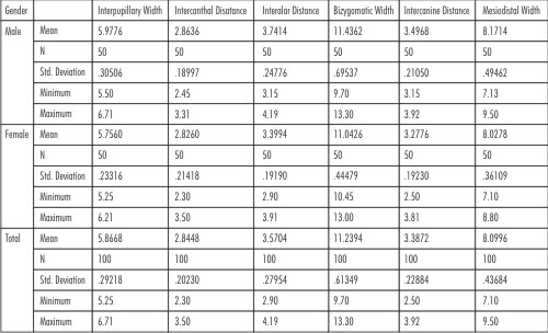 | Table I mean, standard deviation, maximum and minimum values of 100 subjects
 |
The values were greater for men than for women. No significant differences were found between sexes with respect to intercanthal distance.
The paired t-test showed highly significant results in relation to interpupillary width , interalar distance, bizygomatic width, intercanine tip distance. However, intercanthal and mesiodistal width were found to be non significant.(Table II)
 | Table II: Independent t-test
|
The pearson correlation test demonstrated positive correlation between interpupillary width, intercanthal distance, mesiodistal width and interalar and intercanine tip distance . However bizygomatic width was not significantly correlated . The same pattern was observed for both the sexes.(Table III, IV)
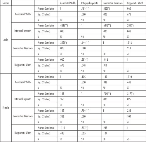 | TABLE III .Pearson correlation (for all subjects and by sex) for MD, IP, IC, BZ
|
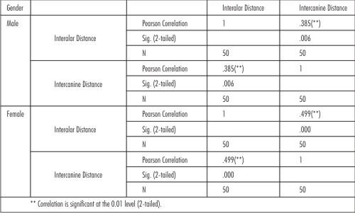 | Table IV: Pearson correlation (for all subjects and by sex) for IA and ICTW
|
The ratios between the mean of interpupillary width, intercanthal distance , bizygomatic width and mesiodistal width of central incisor was 7.3, 3.5, 14.0 respectively in males and 7.1, 3.5, 13.7 respectively in case of females. The ratio between mean of interalar distance and intercanine tip distance was 1.07 in males and 1.03 in females.(Table V)
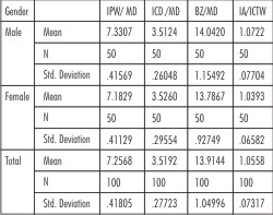 | TABLE V. Ratios between means of different parameters
|
Discussion:
Mesiodistal width of central incisor :
In the present study, all tooth dimensions were significantly larger in men than in women. Gillen et al reported that the maxillary anterior teeth of men were wider and longer than those of women in both white and black populations.4,7 Similarly, Sterrett et al reported the mean width and length of the clinical crowns of the maxillary anterior teeth of men to be significantly greater than the corresponding dimensions in women in a white population.8 In the present study, the mean mesiodistal width of central incisor for men (8.7mm) was significantly greater than the corresponding dimensions for women (8.02mm). These findings are in agreement with the results of related studies.
Bizygomatic width :
Berry has said that the width of maxillary central incisor exists in a ratio of 1:16 to that of the bizygomatic width.9 Later, House and Loop evaluated the ratio published by berry, and found a range of ratios from 1:13 to 1:19, with 1:16 as an average midpoint.10 In the present study the ratio between mesiodistal width of central incisor and the bizygomatic width was found to be 1:14 for males and 1:13.7 for females which is in accordance with the ratios found out by house and loop. The paired t-test performed on the data at 95 percent confidence interval of the difference showed bizygomatic width to be highly significant(p=.001) . However there was no correlation between the bizygomatic width and the central incisor width which is in concurrence with Wazzan et al .
Interpupillary width :
Cesário and Latta showed a mean value of 59.16mm, after measuring 100 subjects of the US Army.11 In the present study the mean of interpupillary width was found to be 59.77 mm in males and 57.56 mm in females, males having greater measurements than females. The mean value for all subjects was 58.66 mm which is in concurrence with Cesário and Latta.
Cesário and Latta showed a mean value of 59.16mm, after measuring 100 subjects of the US Army.11 In the present study the mean of interpupillary width was found to be 59.77 mm in males and 57.56 mm in females, males having greater measurements than females. The mean value for all subjects was 58.66 mm which is in concurrence with Cesário and Latta.
Cesario and Latta found that a ratio of 6.6, which had previously been proposed, existed between the interpupillary distance and the central incisor width in white men and women, and also in black women.Values of 7.7 and 7.5 were found for men and women, respectively by Hasanreisoglu.8 The present study conducted showed a ratio of 7.3 for males and 7.1 for females. Pearson correlation showed positive correlation between interpupillary width and mesiodistal width of maxillary central incisor
Intercanthal Distance:
The ICD showed a median of 28.44 mm, ranging from 23.0 to 35.0 mm . Abdullah found a mean of 28.30 mm and Al Wazzan, measuring the facial segment with a modified Boley gauge, described a range from 25.00 to 39.00mm, with a mean of 31.92mm.12 The different values cited previously possibly could be a result of the ethnic variation of the study sample analyzed. In the present study, the intercanthal distance was the only facial segment that shows no significant difference relative to gender. This finding is in accordance with Laestadius et al and Abdullah et al and Al Wazzan12. The pearson correlation test showed positive correlation between intercanthal distance and mesiodistal width of maxillary central incisor. This finding is in accordance with Gomes et al. These findings indicate that the intercanthal distance could be used to select maxillary anterior teeth for edentulous patients.
Wazzan found a biometric ratio of 1:0.267 between the mesiodistal width of central incisor and intercanthal distance. In the present study, a ratio of 1:0.285 for males and 1:0.284 for females was recorded which is in accordance with the ratio determined by Wazzan.
Interalar width and intercnine tip distance:
The interalar width of the total sample showed a mean of 35.704 mm, ranging from 29.0 mm to 41.9mm . Latta, Weaver, and Conkin found in edentulous patients a mean of 43.93 mm, with a range from 29.00 to 63.00mm. 13
The interalar width was significantly correlated to the mesiodistal width measured on the photo, both between the tips of the maxillary canines.
Mavroskoufis and Ritchie also found the interalar width and the distance between the tips of maxillary canines correlated.14
In the present study the intercanine tip distance ranged between 25.0mm to 39.2mm with a mean of 33.8mm. Hoffman, Bomberg, and Hatch, after measuring the structures by a Boley gauge, found mean values of 34.28 mm for the interalar width, 35.35mm for the distance between the tips of maxillary canines, and 44.85mm for the circumferential arch distance between the distal surfaces.
They concluded that when interalar width was multiplied by 1.31 (or increased by 31%), the calculated value was equal to the circumferential arch distance between the distal surfaces. When the interalar width was multiplied by 1.03 (increased by 3%), the result was equal to the distance between the tips of maxillary canines.
In the present study the ratio between the interalar width and intercanine tip width was 1:1.0558 which is in accordance with Gomes et al who found a ratio of 1:1.03 in Brazilian dentate subjects. The interalar width is highly significant in relation to intercanine tip width. Hoffman, Bomberg, and Hatch points out that although the interalar width and the intercanine tip width are not equivalent, as suggested in previous references in the literature, there is sufficient correlation to use the interalar width increased by 3% as an approximate intercanine tip width when arranging denture teeth.
These findings, together with the results of earlier published reports suggest that methods based on the relationship between the anterior teeth and certain facial measurements can be used as preliminary guides in estimating the width of the maxillary central incisors or the location of the canines in local population. In the present study, limitations such as minor positional differences that can occur during photography might have affected the measurements. Additional research on a greater sample size selected more systematically is needed before extrapolating the results to the general population.
Summary And Conclusion:
Within the limitations of the present study, the following conclusions were drawn:
1. All the facial and dental measurements were greater for men than for women, however no significant differences were found between sexes with respect to intercanthal distance.
2. There was positive correlation between interpupillary width, intercanthal distance and mesiodistal width of central incisor. Interalar distance and intercanine tip distance were also correlated. However bizygomatic width was not significantly correlated. The same pattern was observed for both the sexes.
3. The ratios between the mean of interpupillary width, intercanthal distance , bizygomatic width and mesiodistal width of central incisor was 7.3, 3.5, 14.0 respectively in males and 7.1, 3.5, 13.7 respectively in case of females. The ratio between mean of interalar distance and intercanine tip distance was 1.07 in males and 1.03 in females.
It can be concluded that although various methods of selection of teeth are used, the applicability can vary due to the ethnic differences between populations. Hence these measurements can be used for determining the width and position of the maxillary anterior teeth in local population of Mandi district. In future, studies should be carried out with greater sample size to authenticate the use of these parameters for selection and arrangement of maxillary anterior teeth and make sure that we provide “incredible smiles” with naturalistic looking dentures.
References:
1. Salvatore JE. Esthetics for denture patients. J Prosthet Dent 1980; 44(6):608-613.
2. Gomes Vanderlei Lui. Correlation between Facial Measurements and the Mesiodistal Width of the Maxillary Anterior Teeth. J Esthet Restor Dent 18:196-205, 2006.
3. Isa Z.M., Tawfiq O.F., Noor M.N., Shamsudheen M.I. Regression methods to investigate the relationship between facial measurements and widths of the maxillary anterior teeth. J Prosthet Dent 2010;103(3):182-188.
4. Hasanreisoglu U, Berksun S, Aras K, Arslan I. An analysis of maxillary anterior teeth: facial and dental proportions. J Prosthet Dent 2005; 94:530-8
5. Zarb G.A., Bolender C.L., Hickey J.C., Carlson G.E. Boucher's prosthodontic treatment for edentulous patients (10ed). St. Louis. The C.V. Mosby Co., 1990; pp. 333.
6. Hoffman W, Bomberg TJ, Hatch RA. Interalar width as a guide in denture tooth selection. J Prosthet Dent 1986; 55(2):219-221
7. Gillen RJ, Schwartz RS, Hilton TJ, Evans DB. An analysis of selected normative tooth proportions. Int J Prosthodont 1994;7:410-7.
8. Sterret JD, Oliver T, Robinson F, Fortson W, Knaak B, Russel CM. Width/ length ratios of normal clinical crowns of the maxillary anterior dentition in man. J Clin Periodontol 1999;26:153-7
9. Berry FH. Is the theory of temperaments the foundation of the study of prosthetic art? Dentist's Magazine 1905-1906;1:405
10. House MM, Loop JL. Form and color harmony in the dental art. Whittier (CA): M.M. House; 1939
11. Cesário VA, Latta GH Jr. Relatioship between the mesiodistal width of the maxillary central incisor and interpupillary distance. J Prosthet Dent 1984;52(5):641-3
12. Al Wazzan KA. The relationship between intercanthal dimension and the widths of maxillary anterior teeth. J Prosthet Dent 2001;86:608-12
13. Latta GH Jr., Weaver JR, Conkin JE. The relationship between the width of the mouth, interalar width, bizygomatic width, and interpupillary distance in edentulous patients. J Prosthet Dent 1991;65(2):250-4.
14. Mavroskoufis F, Ritchie GM. Nasal width and incisive papilla as guides for the selection and arrangement of maxillary anterior teeth. J Prosthet Dent 1981;45(6):592-7. |