|
|
|
| Treatment Of Gingival Hyperpigmentation With Diode Laser - An Esthetic Approach |
Anurag Aggarwal 1 , Deepak Bala 2 , Saryu Arora 3 , Manpreet S Walia 4 , Sunil Malhan 5
1 Reader, Deptt of Conservative Dentistry - National Dental College & Hospital, Derabassi
2 Post Graduate Student, Deptt of Periodontology & Oral Implantology - M. M College of Dental Sciences & Research
3 MDS (Senior Lecturer), Deptt. of Prosthodontics - Shri Sukhmani Dental College And Hospital, Derabassi
4 MDS (Professor and Head) Deptt. of Prosthodontics - H.S. Judge institute of Dental Sciences and Hospital, Chandigarh.
5 Professor and Head, Dept of Conservative Dentistry and Endodontics - Desh Bhagat Dental College & Hospital, Mukatsar
|
| Address For Correspondence |
Dr. Anurag Aggarwal
M.D.S. (Senior Lecturer)
Department of Conservative Dentistry
Swami Devi Dayal Hospital and Dental College,
Golpura, Panchkula, India.
Mobile No: +91-9815288381
E mail: anudrrag@gmail.com |
| Abstract |
| Excessive gingival display and gingival hyperpigmentation are major concerns for a large number of patients visiting the dentist. Melanin hyperpigmentation usually does not present a medical problem, but patients usually complain of dark gums as unaesthetic. This problem is aggravated in patients with a "gummy smile" or excessive gingival display while smiling. Esthetic periodontal plastic surgery is especially rewarding in such individuals with compromised esthetics. This case report represents correction of gingival hyperpigmentation by laser ablation using diode laser with satisfactory and pleasant results. |
|
| Keywords |
| Gingival hyperpigmentation, Gingival depigmentation, Repigmentation |
|
| Full Text |
Introduction
In today's dental practice esthetics has become a significant aspect and clinicians are more concerned of achieving acceptable gingival esthetics as well as addressing biologic and functional problems. The color of the gingiva plays an important role in overall esthetics but the principles and the techniques of the management of the problems associated with gingival melanin hyperpigmentation are still not fully established.
Gingival melanin hyperpigmentation is usually encountered in African, Eastasian & Hispanic ethnicity1,2 as well as in certain medical conditions such as endocrine disturbance, Albright's syndrome, malignant melanoma, antimalarial therapy, Peutz-Jeghers syndrome, trauma, hemochromatosis, neurofibromatosis and chronic pulmonary disease3. It varies from individual to individual but directly relates to melanoblastic activity.
Melanin hyperpigmentation usually does not present as a medical problem as it is often physiological rather than pathological but patients may complaint of "black gums" which may pose esthetic problem & embarrassment especially in patients with a ''gummy smile'' or excessive gingival display while smiling or talking. Gingival depigmentation is a periodontal plastic surgical procedure whereby the gingival hyperpigmentation is removed or reduced by various techniques such as mechanical, surgical, chemical, electrosurgical, and cryosurgical techniques but the results are almost similar but not stable for long term4.
Now day's most accepted painless, bloodless, effective, pleasant and reliable technique used for the removal of gingival depigmentation is by mean of diode lasers.
Following case report presents the management of gingival melanin hyperpigmentation with diode laser exclusively for esthetic purposes providing excellent outcome.
Case Report
Patient of age 22 years reported to the department with a chief complaint of having unaesthetic, diffuse, dark-brown to black gingival discoloration in the labial aspect of the maxilla and mandible. Patient requested for cosmetic therapy which will improve the esthetics on smiling. In general the skin pigmentation correlates with gingival pigmentation but in this specific patient the gingival melanin hyperpigmentation was observed that moderately predominated over skin pigmentation. Patient's clinical & medical history revealed physiologic gingival melanin hyperpigmentation. Patient's gingiva was found clinically healthy & free from any visible clinical inflammation thus taking into consideration the patient's chief complaint, laser assisted gingival depigmentation procedure was planned. Whole procedure was explained verbally to the patient in simple and clear language and written informed consent was taken before the procedure. Laser safety precautions were taken during the procedure.
Surgical Procedure:-
The surgical site was anesthetized by local infiltration with 2% lignocaine containing 1:80000 adrenaline.
Laser beam was activated at 2.5 W using brush technique in focused mode i.e. continuous movement of the beam with approximately 20% to 30% overlaps of the laser spots. Lasing time differed according to the degree of pigmentation, the pigmented surface treated, and the epithelial thickness. The beam was initially delivered along the mucogingival junction, moving towards the free gingival margin. The papillary edges and the free gingival margins were initially left untouched to avoid morphological deformations. During lasing, the gingiva was covered with charred layer that was easily removed by wet gauze. A burning smell typical to the procedure was reduced or eliminated by power suction evacuation. No periodontal dressing was given.
The entire gingival surface that required treatment was lased in a single session.
Patient was instructed to avoid trauma to the treated gingiva and refrain from acidic and hot food for one week.
The patient was reevaluated 10 days and 3 months after the last lasing session. Evaluation included clinical examination and comparative clinical photographs (Fig 1-5).
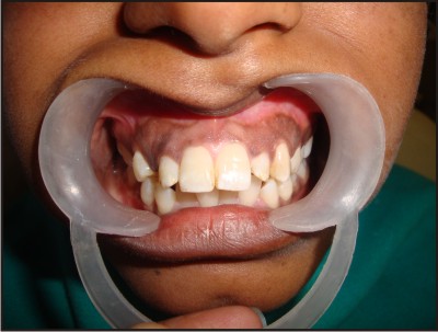 | Pre Operation
 |
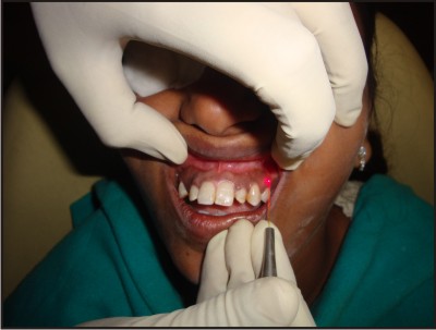 | During Surgery
|
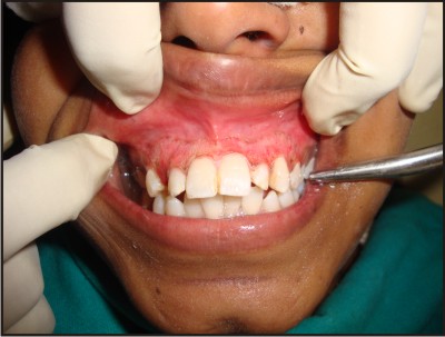 | Post Operation
|
 | 3 Month Post Operation
|
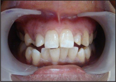 | 1 Year Post Operation
|
Results
No discomfort, teeth sensitivity, pain or bleeding complications were found intra or postoperatively. Therefore no antibiotics or analgesics were prescribed. The results were very pleasing and the gingival appearance was satisfactory 2 to 4 weeks after treatment. Patient did not needed any rest or experienced any interruption in performing normal activities. During the 1 year follow-up, no signs of repigmentation were observed.
Discussion
Recently, laser ablation has been recognized as an effective, pleasant and reliable technique. It is preferred over other techniques by many clinicians. When laser energy interacts with biologic tissue, the effect is influenced by the emitted wavelength, laser energy, and time of exposure and rate of movement of the fiber tip across the target tissue. Actually the absorption of laser energy in the tissue is the key element of laser-tissue interaction. Pain reduction, intra- and postoperatively, and rapid wound healing are important advantages of laser use5. In this present case report, patient reported absence of pain intraoperatively, postoperatively which may be due to the sealing of the ends of the sensory nerve 6.
Here the brush technique with a defocused beam was used as used by Tal H et al 2003 which seems to be safe and effective7. However Atsawasuwan et al 2000 described gingival fenestration resulting in bone exposure up to 4 weeks with a focused Nd:YAG laser beam using the brush technique after the procedure. Although the fenestration healed uneventfully with the defocused beam, it was more time-consuming8.
In the present case report no repigmentation occurred in treated patient till one year follow up.
Conclusion
Present case report concluded that the management of gingival melanin hyperpigmentation with diode laser is a safe and effective procedure providing excellent esthetic outcome at one year follow up. The results were pleasing to the patient which is the ultimate goal of any therapy. Following laser ablation the gingiva healed uneventfully and completely regenerated with no infection, pain, swelling, or scarring. So to conclude that depigmentation with laser can be used as successful procedure.
References
1. Fry L, Almyeda JR. The incidence of buccal pigmentation in caucasoids and negroids in Britain. Brit J Dermatol 1968; 80(4): 244-7.
2. Tamizi M, Taheri M. Treatment of severe physiologic gingival pigmentation with free gingival autograft. Quintessence Int 1996; 27(8): 555-60.
3. Leston JM et al. Oral mucosa: variation from normalicy, part II. Cutis 2002; 69(3): 215-7.
4. Roshna T, Nandakumar K. Anterior esthetic gingival depigmentation and crown lengthening: Report of a case. J Contemp Dent Pract 2005; 15: 139-47.
5. Ishikawa I, Aoki A, Takasaki AA. Potential applications of Erbium:YAG laser in periodontics. J Periodontal Res 2004; 39: 275-85.
6. Schuller DE. Use of the laser in the oral cavity. Otolaryngol Clin North Am 1990; 23: 31-42.
7. Tal H, Oegiesser D, Tal M. Gingival Depigmentation by Erbium: YAG Laser: Clinical Observations and Patient Responses. J Periodontol 2003; 74: 1660-7.
8. Atsawasuwan P, Greethong K, Nimmanon V. Treatment of gingival hyperpigmentation for esthetic purposes by Nd: YAG laser: Report of 4 cases. J Periodontol 2000; 71: 315-21. |
|
|
|
|
|
|