Introduction
Lupus erythematosus (LE) is a connective tissue disease including Discoid Lupus Erythematosus (DLE) and Systemic lupus erythematosus (SLE)1. Discoid lupus erythematosus, less aggressive form of LE is a chronic, scarring, atrophy producing photosensitive dermatosis. It rarely (5%) progresses to SLE. It is most commonly seen in middle aged women. The clinical manifestations include the appearance of discoid lesions solely on the skin, most commonly on the face, scalp, oral mucous membrane, chest, back and extremities2. Systemic lupus erythematosus is a clinically heterogeneous, autoimmune disease characterized by the production of auto antibodies. It usually occurs in the postmenopausal females. It has got a multisystem clinical presentation with the involvement of dermatological, hematological, renal, neural, and oral involvement. Oral lesions can be either in the form of discoid lesions or ulcers. 1 Extremely vigilant examination and necessary investigations are required for the accurate diagnosis. Steroids form the main line of treatment.
Case Report
A 40 year female patient came to the department with the chief complaint of ulcers in the oral cavity since 15 days. History of present illness revealed that the ulcers started 15 days back as tiny pin point ulcers on the lower labial mucosa and left buccal mucosa. They gradually increased in size and involved the upper and lower labial mucosa, hard palate, and left buccal mucosa. She visited dentist where she was advised Nitragel for the local application on the same. The lesions aggravated on the application of medication. They were extremely painful and associated burning sensation was present even on taking normal foods. Her past medical history revealed that she was asthmatic since 16 years and was on medication - Asthalin inhaler (salbutamol-100mg). She used to take one to two puffs per day according to the need. She had skin lesions one year back which started as erythmatous rashes on the face and gradually involving the back and lower part of neck. She visited a dermatologist and was advised Tab .Kenacort (4mg) and Clobetasolgel (0.05%). She also gave the history of blood transfusion on three separate occasions since one year. She complained of joint pains especially knee & ankle joints pains for which she had visited a specialist and was found to be Rheumatoid factor negative. Her menstrual history was normal. General physical examination of the patient revealed a moderately build and nourished patient with all vital signs within the normal limits. Conjunctivalpallor was evident.Lymph Node examinationrevealed solitary, bilateral submandibular lymph nodes measuring approx 1.5 X 1.5 cms in size, oval in shape, soft in consistency, tender and palpable.
Extra oral examination revealed diffuse brownish black pigmentation involving the malar region bilaterally involving the nasolabial fold region, whole length of the nose, philtrum region, and chin and extending till the upper part of the neck. It was interspersed with isolated areas of depigmentation and annular erythmatous areas. Similar lesions were evident in the mid clavicular region and also the whole back region. The lesions were non tender on palpation. Fissuring was evident bilaterally at the corner of mouth. Blood encrustations were evident involving the vermillion zone region of the upper and the lower lip. There was loss of distinction of the vermillion border of the lower lip. On palpation the lesions were extremely tender and bled on minor provocation. Intraorally bilaterally diffuse erythematous lesions were present on the right and the left buccal mucosa. Diffuse erythematous lesions were evident involving the entire palate. They were interspersed with few white keratotic lacy lesions. The lesions were extremely painful. Gingival inflammation was present. Hard tissue examination revealed full complement of maxillary and mandibular dentition.
Considering the history, mode of onset of the lesions, clinical appearance of the lesions we arrived at the provisional diagnosis of oral manifestation of systemic lupus erythematosus. The differential diagnosis ofErythema multiformae, Stomatitis veneneta and Pemphigus vulgaris were considered. Hemogram revealed reduced values of hemoglobin, white blood cells and platelet counts. ESR was raised. Peripheral blood smear report revealed microcytic hypochromic anemia with leucopenia and thrombocytopenia. Random blood sugar levels were within the normal limits. Blood urea was 40 mg% and albumin was detected in the urine examination.Pulmonary function tests revealed pleuritis and severe chest congestion. PEFR (peak expiratory flow rate) was reduced. Anti-nuclear antibodies (ANA) were negative however LE cell preparation results was positive ,hence a final diagnosis of Systemic lupus erythematosus was made.
The treatment modality involved a regimen of drugs for the specific disease condition as well as for the accompanied symptoms after consultation with the physician. The regimen for this particular patient included -
Benzydyamine Hydrochloride 0.15% thrice daily,
Triamcinolone acetonide-0.1 %- for local application thrice daily.
Cefadroxil250 mg b.i.d for 5 days
60 million spores of lactobacillusb.i.d for 10 days
I.V Deriphylline 12th hourly for 5 days
Inj Frusemide-10mg for 15 days
Dexamethasone sodium and neomycin sulphate(0.5%) topical application
InjDexona 4mg for 15 days
Ranitidine 150 mgo.d. for 7 days.
After 15 days, the patient reported marked improvement in the signs and symptoms. There was significant reduction in the buccal mucosal lesions. The skin lesions also showed signs of remission. She was advised to continue the intraoral topical medication as well as topical application for the skin surface. After 1 month the patient reported with complete healing of the palatal lesions. Mild erythematous lesions were evident on the lower labial mucosa and right and left buccal mucosa. There was significant reduction in the burning sensation during her second visit after 1.5months. Patient is still under follow up.
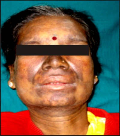
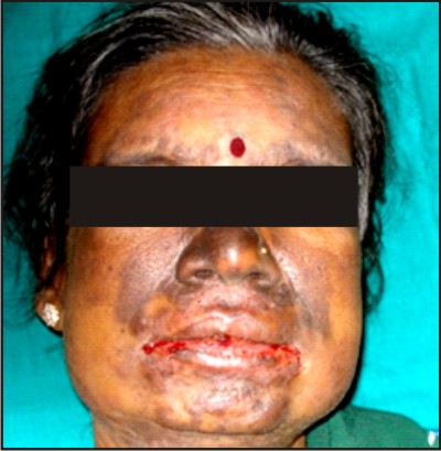 | Figure 1- Frontal View Of The Patient
 |
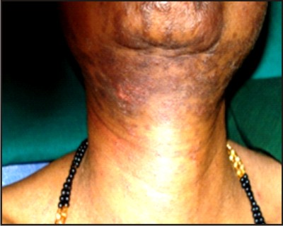 | Figure 2- Lesions Involving The Chin And Upper Part Of Neck.
|
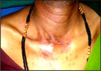 | Figure 3- Lesions Involving The Midclavicular Region
|
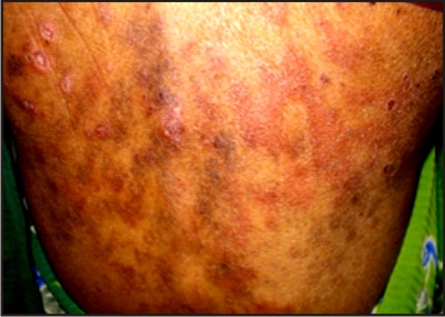 | Figure 4 - Lesions Involving The Back Region
|
 | Figure 5 - Lesions Involving The Lips.
|
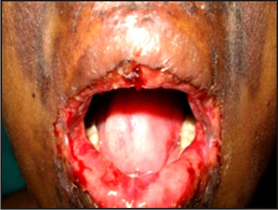
 | Figure 6 - Lesions Involving The Palate
|
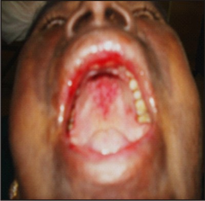
 | Figure 7 - Follow Up After 15 Days Frontal View Of The Patient
|
 | Figure 8 - Follow Up After 1 Month Healed Lesion Involving Lower Lip And Mucosa.
|
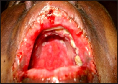
 | Figure 9 - Follow Up After 1 Month Healed Lesions On The Palate
|
Discussion
SLE is a chronic multifactorial prototypic autoimmune disease characterized by the presence of auto antibodies directed against nuclear antigens.This disease commonly affects young women of child bearing age with aFemale: Male ratio of 12:1 within the age group of 15-45yr and ratio of 2:1 inchildren or elderly3.
The clinical heterogeneity of this disease is mirrored by its complex etiopathogenesis. In presence of appropriate antigens SLE develops due to formation of soluble immune complexes mainly composed of IgG& IgM4 following a pattern of type III hypersentivity reaction triggered by endogenous antigens which can be generalised or organ specific. Because of the size of the immune complexes &its known affinity organs like kidneys, lungs, joints are the target sites5.
Tissue damage is caused primarily by platelets & neutrophils, lesions contain neutrophils, immune complex deposits & complement C3a,C4a ,C5a6. Later stages have macrophage infiltrations which are involved in healing5.
Genetic factors & specific genetic loci (HLA-DR, HLA-DQ, C-RP) are also important for the pathogenesis of SLE4. Environmental factors like exposure to U-V light (photosensitivity), drugs (pharmacogenetics) & infections ( with Epstein barr virus) also precipitate the development of SLE7.
Clinical features of SLE are diverse and include1
Constitution Features like fatigue, weight loss, fever etc.
Renal Diseases or Lupus Nephritis affects 30% of the patients and is the most dangerous, life threatening complication.
Neuropsychiatric Lupus includes headache, depressions, seizures and psychosis. Migraines are also more prevalent in these patients.
Musculoskeletal Disease includes - Myalgia & Arthralgia.Arthralgia is asymmetric and migratory is often the earliest manifestation. The joints of the hands are most often affected. The arthritis is moderately painful and nondestructive. Deformities observed are usually due to tendon inflammation (Jaccoud'sArthropathy), rather than degeneration.
Dermal involvement in lupus includes malar and discoid rashes and generalized photosentivity. Alopecia also results due to hair follicle plugging with keratin Oral lesions such as desquamative gingivitis, marginal gingivitis or erosive mucosal lesions have been reported in up to 40% of patients. Patients with advanced cases of SLE may have features of Sjögren's syndrome, such as dry eyes, mouth and skin.
Haematological features include normocytic normochromic anaemia, thrombocytopaenia (sometimes, but notalways associated with antiphospholipid antibodies) andleukopaenia. Pleuritis, causing chest pain, cough and breathlessness is the most common pulmonary manifestation of SLE8. Pleural effusions and parenchymal damage often lead to pneumonitis. Subsequently, a hospitalized patient with SLE would be at great risk of acquiring pneumonia.9 Chest pain warrants a detailed workup to rule out pulmonary embolism and myocardial infarction, especially in those who test positive for antiphospholipid antibodies.7 Two well-known cardiac features of SLE are pericarditis and endocarditis. Patients with SLE are at increased risk of atherosclerosis. Chronic inflammation and the use of corticosteroids contribute to this risk. Gastrointestinal involvement 1 most commonly results in non-specific abdominal pain and dyspepsia Hepato-splenomegaly can come and go with disease activity. Mesenteric vasculitis is very rare, but can be life threatening, especially if it leads to perforation.
Various vascular manifestations-Raynaud's phenomenon, occur causing the classical triphasic colour change.
The clinical differential diagnosis includes Apthous ulcers, Erythema multiformae, Lichen planus, Stomatitis venenata, Stomatitis medicamentosa, Seborrheic dermatitis, actinic keratosis and fixed drug eruptions5. Diagnosis is based on clinical examination, laboratory examination and Immunological tests. In 1997 American College of Rheumatology gave the diagnostic criteria for SLE in which 4 out of 11 symptoms occurring either serially or in succession confirm SLE. These are 5
Malar Rash
Discoid rash
Photosensitivity
Oral Ulcers
Arthritis
Serositis
Renal Disorder
Neurological Disorder
Hematological Disorder
Immunological Disorder
Antinuclear Antibody
If a diagnosis of SLE is suspected, then the most useful preliminary testing includes a complete blood count with differential white blood cell counts. This count will reveal chronic anemia that is normocytic-normochromic with thrombocytopenia and lymphocytopenia.6,7
Other disease-specific tests for autoantibodies like antinuclear antibodies,anti-double stranded-DNA, anti-Smith antibody, anti RO-(SSA), anti-phospholipidantibody,and complement, C3, C4levels1.
Treatment of SLE is based on prevention, reversal of inflammation, maintaining states of re-mission and alleviation of symptoms.Treatment plan includesthe use of protective sunglasses, protective clothing and Sunscreen (SPF>15)5.Various medicinal therapies include- Non-steroidal anti-inflammatory drugs, cyclooxygenase-2 selective inhibitors and antimalarialswhich are generally effective for musculoskeletal complaints and mild serositis5,10. Systemic corticosteroids, such as prednisone, are reserved for patients with morbid symptoms associated with significant organ involvement, particularly renal, central nervous system and systemic vascular diseases10. The dosage of corticosteroid is progressively tapered as signs and symptoms resolve. However, some patients may require a maintenance dose to remain in remission.10
Perioperative Management by the Dentist 5,9,1
Dentists must enforce preventive dental care and monitor patients with SLE closely for head and neck infections because they are predisposed to severe infections. These infections are often silent and difficult to detect because of anabsence of pain and swelling.Thorough clinical examination is required to avoid overlooking infections. Infections can progress rapidly in patients with SLE because of disease or therapy-related immunosuppression. Most patients with SLE can have a superimposed antiphospholipid antibody syndrome that predisposes them to thromboembolic events, such as arterial and venous thrombosis, pulmonary embolism, stroke and myocardial infarction9. It is therefore important to document whether these patients are managed with anticoagulation therapy, aspirin or warfarin before dental surgery. Recent laboratory tests may be indicated preoperatively to determine platelet count, prothrombin time and the international normalized ratio (INR) for blood clotting time. Local measures for maintaining hemostasis may also be required. Antibiotic prophylaxis before bacteremia-associated dental and oral surgical interventions is required to prevent infectious endocarditis in SLE patients with valvular damage1.
Chronic renal failure because of SLE will influence the choice or dosage of medications prescribed by dentist .Patients suffering from chronic renal failure is often on dialysis. Dental surgery should be planned one day after dialysis treatment to ensure elimination of adminis-tered medications and their by-products1.
For patients with neuropsychiatric symptoms the dentist's role is paramount to rule out odontogenic, temporomandibular joint and associated myofascial sources of pain1. Patients on long-term corticosteroids may require supplemental dosing on the day of a potentially stressful dentoalveolar surgery5.
Conclusion
Lupus is like a puzzle, with genetics, gender, and the environment being important pieces of the puzzle which when fit together result in this autoimmune malady. A multidisciplinary approach to medical consultation and appropriate referrals ensures comprehensive medical and dental management of patients with SLE. (1982)
References
1. Manson JJ, RahmanA . Systemic Lupus Erythematosus. Orphanet J Rare Dis. 2006; 1:6.
2. ChandraSekhar .P, Suvarna M, ArvindBabu.R.S , Anuradha.C , Shamala R., Reddy .B.V.R- Lupus erythematosus - A report of 3 cases, J OrofacSci, 2010; 2: 30-35.
3. Susan M. Lupus Update: Perspective and Clinical Pearls. Cleveland Clinic Journal of Medicine 2009; 76: 137-42.
4. Nath SK, Kilpatrick J, Harley JB. Genetics of human systemic lupus erythematosus: the emerging picture. CurrOpinImmunol2004; 16(6):794-800.
5. Albilia JB, Lam DK,Clokie CML, Sándor CKB, Systemic Lupus Erythematosus: A Review for Dentists. JCDA, 2007; 73:823-828
6. Cervera R, Khamashta MA, Font J, Sebastiani GD, Gil A, Lavilla P, et al. Systemic lupus erythematosus: clinical and immunologic patterns of disease expression in a cohort of 1,000 patients. The European Working Party and Systemic Lupus Erythematosus. Medicine (Baltimore) 1993; 72:113-24.
7. Estes D, Christian CL. The natural history of systemic lupus erythematosus by prospective analysis. Medicine (Baltimore) 1971; 50: 85-95.
8. Paran D, Fireman E, Elkayam O: Pulmonary disease in systemic lupus erythematosus and the antiphospholpid syndrome. Autoimmun Rev 2004, 3:70-75.
9. Fessler BJ, Boumpas DT. Severe major organ involvement in systemic lupus erythematosus. Diagnosis and management. Rheum Dis Clin North Am 1995; 21; 1:81-98.
10. Fessler BJ, Alarcon GS, McGwin G Jr, Roseman J, Bastian HM, Friedman AW, et al. Systemic lupus erythematosus in three ethnic groups: XVI. Association of hydroxychloroquine use with reduced risk of damage accrual. Arthritis Rheum 2005; 52; 5:1473-80. |