|
|
|
| Gingival Depigmentation In Children : A Case Report |
Manjul Mehra 1 , Rashu Grover 2 , Sunil Gupta 3 , Gunmeen Sadana 4
1 Senior Lecturer, Dept Of Pedodontics And Preventive Dentistry - Sri Guru Ram Dass Institue Of Dental Sciences And Research, Amritsar
2 Senior Lecturer, Dept Of Pedodontics And Preventive Dentistry - Sri Guru Ram Dass Institue Of Dental Sciences And Research, Amritsar
3 Professor, Dept Of Pedodontics And Preventive Dentistry - Sri Guru Ram Dass Institue Of Dental Sciences And Research, Amritsar
4 Professor And Head, Dept Of Pedodontics And Preventive Dentistry - Sri Guru Ram Dass Institue Of Dental Sciences And Research, Amritsar
|
| Address For Correspondence |
Dr. Manjul Mehra(M.D.S.) Senior Lecturer,
Dept Of Pedodontics And Preventive Dentistry,
Sri Guru Ram Dass Institue Of
Dental Sciences And Research,
Amritsar, 143001 Punjab India
Phone Number : 08146133366
E-mail Address : mehramanjul@yahoo.co.in |
| Abstract |
| In recent years, there is an increasing need for esthetics and growing cosmetic demands for a pleasing smile in many individuals. In particular, parents are more conscious of the black or dark pigmentation patches on the facial aspects of the gingiva of their child, which may be strikingly apparent during smiling and speaking. Till date very little literature has been published regarding clinical methods of treatment of pigmented gingiva in children. A case is reported here in which a simple and effective surgical depigmentation was performed without the use of any sophisticated instruments or apparatus. |
|
| Keywords |
| Glove type finger prosthesis,Silicone,Amputee |
|
| Full Text |
Introduction
Hyperpigmentation of the gingiva is caused by excessive melanin deposition by the melanocytes mainly located in the basal and suprabasal cell layers of the epithelium [1]. Several local and systemic factors cause melanin pigmentation, including physiological or racial pigmentation, smokers melanosis, pigmented nevus, melanotic macula, Addison disease, Peuutz-Jeghers syndrome, HIV infection and drugs such as minocycline and anti- malarial drugs[2]. It has been observed that there is positive correlation between gingival pigmentation in children and parental smoking, this pigmentation may be induced by the stimulation of melanocytes by stimuli present in tobacco smoke such as nicotine and benzopyrene [3].
The hyperpigmentation of the gingiva is benign in most cases, and is not a medical concern. However, it may cause esthetic problems for some individuals, especially those who have gummy smile[4]. Gingival depigmentation is a periodontal plastic surgical procedure whereby the gingival hyperpigmentation is removed or reduced by various techniques. The first and foremost indication for depigmentation is patient demand for improved esthetics.
The present case report, describes a simple and effective surgical depigmentation technique that does not require sophisticated instruments or apparatus yet yields esthetically acceptable results along with patient's satisfaction.
Case report
A 9-year-old male child visited Department of Pedodontics, Sri Guru Ram Das Institute of Dental Sciences And Research, along with his parents with the concern of his unaesthetic anterior gingival pigmentation and his malalligned teeth. On examination, the patient had a very high smile line that revealed the deeply pigmented gingiva on the labial surface of both maxillary and mandibular arches. The color of his gingiva was dark to black (Fig. 1).
 | Fig 1 : Pre-operative photograph showing pigmented gingiva
 |
The patient had a mixed dentition period with maxillary right central incisor in cross bite. Depigmentation procedure was planned only in maxillary arch along with anterior inclined plane for correction of cross bite.
A scalpel surgery with bur abrasion was planned to perform the depigmentation. Following the administration of local anaesthetic solution, A Bard Parker handle with a No.15 blade and a high speed hand piece with diamond bur were used to remove the pigmented layer (Fig. 2).
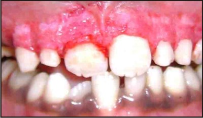 | Fig.2 : Immediately after surgery of the maxillary anterior
|
Pressure was applied with sterile gauze soaked in local anesthetic agent to control hemorrhage during the procedure. After removing the entire pigmented epithelium along with a thin layer of connective tissue with the scalpel, abrasion with diamond bur was done to get the physiological contour of the gingiva, the exposed surface was irrigated with saline. It is recommended to use the largest size diamond bur(diameter of ball 2mm or 2.5mm) because small burs do not smoothen surfaces easily and have a tendency to make small pits in the area to be corrected. Care must be exercised to use feather-light brushing strokes to remove the pigmented areas without holding the bur in one place. All the remnants of the melanin pigment or pigmented areas of the epithelium should be completely removed to prevent possible relapse of the problem. Surgical area was covered with a periodontal pack and post-operative instructions were given. Analgesic was prescribed for the management of pain. After one week, the pack was removed and the surgical area was examined. The healing was uneventful without any post surgical complications. The gingiva became pink and healthy within 5 weeks after ablation. At 9 month follow up, there was no recurrence of gingival hyperpigmentation (Fig 3). The patient and his parents were very impressed with such a pleasing aesthetic outcome.
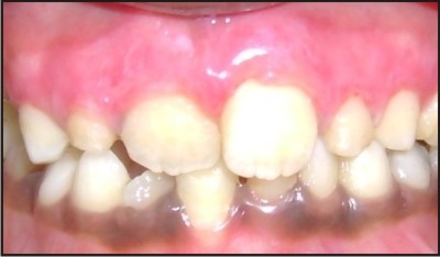 | Fig.3 : 9 month post operative showing pink and firm gingiva
|
Discussion
In recent years, there is an increasing need for esthetics and growing cosmetic demands for a pleasing smile in many individuals. In particular, parents are more conscious of the black or dark pigmentation patches on the facial aspects of the gingiva of their child, which may be strikingly apparent during smiling and speaking. Till date very little literature has been published regarding clinical methods of treatment of pigmented gingiva in children. Different treatment modalities have been used for this procedure in adults. The selection of a technique for depigmentation of the gingiva should be based on clinical experience, patient's affordability and individual preferences.
Electrosurgery requires more expertise than scalpel surgery. Prolonged or repeated application of current to tissue induces heat accumulation and undesired tissue destruction [5]
Cryotherapy is a method of tissue destruction by rapid freezing. The cytoplasm of the cells freeze leading to denaturation of proteins and cell death. This procedure does not require the use of local anesthesia, is relatively a painless procedure and has shown to produce excellent results.However it is followed by considerable swelling and it is also accompanied by increased soft tissue destruction as the depth of penetration cannot be controlled [6].
Recently, a laser has been used to ablate cells containing and producing the melanin pigment. This has the advantages of easy handling, short treatment time, hemostasis and decontamination and sterilization effects .The Nd:YAG laser produces invisible, near-infrared light with a wavelength of 1,064 nm. Because the Nd:YAG laser has rays that have an affinity for melanin or other dark pigments, it works more efficiently when the beam is applied under the presence of a pigment . No significant side effects of scarring, or textural or pigmentary changes have been reported and the incidence of hypopigmentation has been reported as lower than that by other types of lasers[7]. Hyuj-Jin et al found both the Nd:YAG laser and the high speed rotary instrument seem to be effective in the esthetic treatment of gingival melanin hyperpigmentation[8]. G Berk et al pointed out that Er,Cr:YSCG Laser was a good and safe choice for removal of pigmented gingiva without local anesthesia[9]. But this approach needs expensive and sophisticated equipment that is not available commonly at all places and makes the treatment very expensive .
Free gingival grafting is quite an invasive and extensive procedure and has not been advised for depigmentation procedures routinely. but it has the disadvantage of a second surgical site, additional discomfort and poor tissue color matching at the recipient site [11].
Bone denudation procedure is again an invasive method not used for the obvious reasons of bone loss and the discomfort involved in the procedure for the patient. Gingival depigmentation has been attempted by displacing the flap (push back technique), by Kon et al and have reported that melanocytes may lose their ability transiently to produce and transfer the pigment to the keratinocytes, but return to normal much faster than do melanocytes observed after gingivectomy or other procedures[12]
Among the mentioned techniques, we found the scalpel and abrasion technique relatively simple and versatile and it required minimum time and effort. No sophisticated and expensive armamentarium were required, only blade and bur were sufficient. The procedure essentially involves surgical removal of gingival epithelium along with a layer of the underlying connective tissue and allowing the denuded connective tissue to heal by secondary intention. The new epithelium that forms is devoid of melanin pigmentation . However, scalpel surgery may cause unpleasant bleeding during and after the operation, and it is necessary to cover the exposed lamina propria with periodontal dressing for 7 to 10 days [13].
Though the initial result of the depigmentation surgery is highly encouraging, repigmentation is a common problem. The exact mechanism of repigmentation is not understand, but according to migration theory, active melanocytes from the adjacent pigmented tissue migrate to treated areas, causing repigmentation [14].Dummett and Bolden observed partial recurrence of hyperpigmentation in 6 out of 8 patients after gingivectomy at 1 to 4 months[1],where as Permutter and Tal et al did not observe repigmentation until 20 months after cryosurgical depigmentation[15] .No recurrence of hyperpigmentation was found in any of the 4 patients treated by Atsawasuwan et al at 11 to 13 months after gingival depigmentation using Nd:YAG Laser[10] . Sameer A .Mokeem observed no repigmentation occurring in any of the three patients treated with surgical abrasion after 18 months[16].
In the present case, repigmentation was not observed during a short follow up period (9 month). However, long -term observation are required to determine the efficacy of depigmentation in children. In future, even if gingival repigmentation occurs in this patient, the same procedure could be repeated in the same region.
The timing of doing depigmentation procedure in children is not clear in the literature. However, personal experience has found that delay is not necessary as the children are conscious about their dental esthetic appearance and that of the other children.
The depigmentation procedure was successful and both the patient and his parents were satisfied with the result and most important ,his self esteem has improved . Thus, we conclude that depigmentation of hyperpigmented gingiva by scalpel surgery with bur abrasion is simple, easy to perform, cost effective and above all provides minimum discomfort to the patient with esthetically pleasing results.
References
1. Dummett CO. Overview of normal oral pigmentations, J Indiana Dent Assoc 1980;59(3):13-18.
2. Cicek Y, Ertas U. The normal and pathological pigmentation of oral mucous membrane: a review. J Contemp Dent Pract 2003 Aug 15;4(3):76-86.
3. Hanioka T, Tanaka K, Ojima M, Yuuki K. Association of melanin pigmentation in the gingiva of children with parents who smoke. Pediatrics 2005 Aug;116(2):186-90.
4. Hoexter DL. Periodontal aesthetics to enhance a smile. Dent Today 1999;18(5): 78-81.
5. Gnanasekhar JD, Al Duwairi YS. Elecrosurgery in Dentistry. Quintessence Int 1998;29:649-54.
6. Yeh CJ. Cryosurgical treatment of melanin-pigmented gingiva. Oral Surg Oral Med Oral Pathol Oral Radiol Endod 1998;86:660-3.
7. Goldstein A, White JM, Pick RM. Clinical applications of the Nd:YAG laser. In: Miserendino L, Pick RM, editors. Lasers in dentistry. Chicago: Quintessence Publishing Co.; 1995. p. 199-216.
8. Hyuk-jin Ko,Jin-Woo Park ,Jo-Young Suh.Esthetic treatment of gingival melanin hyperpigmentation with a Nd:YAG Laser and high speed rotary instrument:comparative case report.J Periodontal Implant Sci 2010;40:201-205.
9. G.Berk, K.Atici, N.Berk : Treatment of Gingival Pigmentation with Er,Cr:YSGG Laser.J Oral Laser Application 2009;5:249-253.
10. Atsawasuwan P, Greethong K, Nimmanon V. Treatment of Gingival hyperpigmentation for esthetic purposes by Nd: YAG laser: Report of 4 cases. J Periodontol 2000;71:315- 321.
11. Tamizi M, Taheri M. Treatment os severe physiologic gingival pigmentation with free gingival autograft. Quintessence Int. 1996;27(8):555-8.
12. Kon S, Bergamaschi 0. Dome Al. Ruben MP. Melanin Repigmentation after Gingivectomy: A 5-Year Clinical and Transmission Electron Microscopic Study in Humans. Int J. Periodont RestDent,13:85-92,1993.
13. Almas K, Sadiq W: Surgical Treatment of Melanin- Pigmented Gingiva: An Esthetic Approach. Indian Journal of Dental Research, 2002; 13( 2): 70-73.
14. Begamaschi O, Kon S, Doine AI, Ruben MP. Melanin repigmentation after gingivectomy: A five year clinical and transmission Electron Microscopic Study in Humans. Int Journal of Periodontics & Restorative Dentistry, 1993;13(1):85-92
15. Perimutter S.Tai H ,Repigmentation of the gingiva following. Injury.JPeriodontology 1986;57:48-50
16. Sameer A .Mokeem .Management of gingival hyperpigmentation by surgical abrasion -Report of three cases .Saudi dental Journal 2006;18:3:162-165. |
|
|
|
|
|
|