Introduction
Improvements in procedural techniques in endodontics have led to saviour of even badly mutilated teeth. However, the very purpose of retaining teeth by endodontic treatment is defeated if such teeth do not fulfil the objective of active function. Restoration of endodontically treated teeth commonly requires the partial removal of obturating material in the canal in order to prepare the post space, and this procedure can affect the apical seal (1). Various methods have been put forward from time to time. Almost all methods are designed keeping in mind the maintenance of three dimensional seal attained at the apex of such teeth. Various concerns addressed during removal of gutta percha are post length, method and the time of removal of gutta percha (2, 3, 4). Removal of gutta percha can be accomplished by use of thermal, mechanical or chemical means. Literature reveals that no method employed for removal gives the ideal results and there is always a likely hood of disturbing the apical seal which may lead to failure (4, 5). Integrity of apical seal also depends a lot on the time of removal of obturating material from the coronal part of the root canal (6, 7, 8, 9 ). This study was undertaken to compare and quantitatively evaluate two frequently employed obturating techniques for their effectiveness in maintaining integrity of apical seal after post space preparation at varied time intervals.
Material and methods
One hundred extracted permanent maxillary anterior were selected for the study. Only those teeth were selected which were caries free and required extraction due to periodontal reasons. The crown of tooth and if required, a part of root was sectioned to standardize the length of all the samples to fifteen millimetres. All root canals were prepared till size 40 at apex. After instrumentation a No. 15 Reamer was passed through the apex to ensure the apical patency.
Forty five samples were obturated by lateral condensation technique using Gutta percha points and zinc oxide eugenol cement sealer (Group A). Forty five samples were obturated by sectional method maintaining a standard length of five millimetres of gutta percha cone in each tooth. In this group the same zinc oxide eugenol sealer was used. These samples were coated with two layers of enamel paint, leaving only one millimetre of apex without it (Group B). Five samples were used as positive control where no obturation was done and the samples were coated with two layers of enamel paint, leaving only one millimetre of apex without it. Five samples acted as negative control and in these no obturation was done but their apices were closed with two layers of enamel paint. All samples were stored in 100% humidity at 37°C.
For Group A samples in which lateral condensation technique was employed, a heated endodontic plugger was used to remove gutta percha from coronal part of the root canal so that five millimetre of gutta percha filling was left in the apical part. Peeso reamers were used for preparing post space. These samples were divided into three subgroups
Subgroup A1 - Gutta percha filling was removed and post space prepared 2 days after obturation
Subgroup A2 - Gutta percha filling was removed and post space prepared 7 days after obturation
Subgroup A3 - Gutta percha filling was removed and post space prepared 14 days after obturation
For Group B samples in which sectional technique was used, only post space preparation was with peeso reamers was done and these were subdivided into three subgroups in a similar way to Group A-
Subgroup B1 - Post space prepared 2 days after obturation
Subgroup B2 - Post space prepared 7 days after obturation
Subgroup B3 - Post space prepared 14 days after obturation
Dye penetration analysis- Apical 5 millimetre of all the samples was immersed in 2% aqueous solution of Methylene blue dye . The samples immersed in the dye were stored in an incubator at 37°C for 14 days. Afterwards samples were dissolved in 10 millilitres 50% Nitric acid and the solution was analysed using spectrophotometer for optical density. The readings obtained were tabulated and put to statistical analysis.
Results
The study was conducted to evaluate the effect of post space preparation on apical seal of endodontically treated teeth leaving intact 5 mm of apical gutta percha. One hundred maxillary anterior teeth were selected for the study. The results obtained have been tabulated in Table-1 and Table- II. The mean optical density for negative control was 0.02 and for positive control it was 0.988(Table III). These readings were taken as extreme values for both the groups. Samples showing optical density values lower than 0.02 and higher than 0.988 were discarded (Table IV).
The readings obtained for each group were tabulated (Table I & II) and were put to statistical analysis, employing two way analysis of variance.
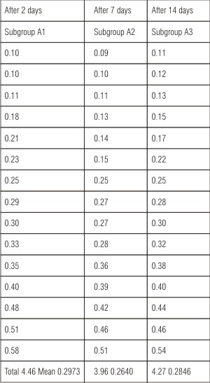 | Table-I Apical Optical Density In Group-A (Lateral Condensation Technique)
 |
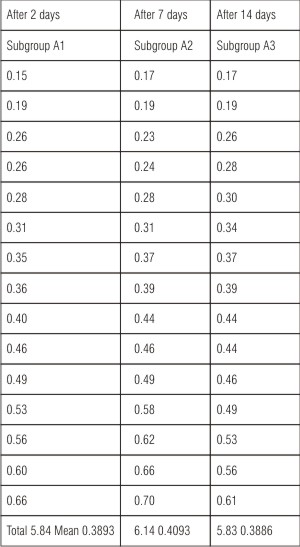 | Table-II Apical Optical Density In Group-B (Sectional Obturation Technique)
|
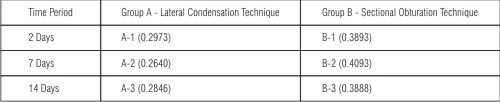 | Table-III Comparison of Group A and Group B
|
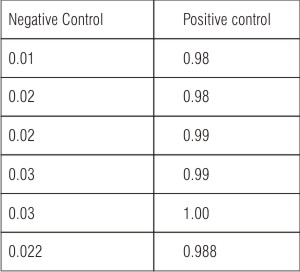 | Table-IV Optical Density Mean Value
|
The null hypothesis assumed was:
1. Method of obturation of root canal had no significant effect on apical leakage.
2. Time of post preparation had no effect on apical leakage.
The sample evidence showed insignificant F value 0.36 (Table V) as regard to second null hypothesis. It indicates that the timing of post preparation had no significantly different effect on apical leakage.
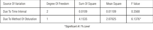 | Table-V Two Way Analysis Of Variance(Anova)
|
The F value in case of first null hypothesis was 6.1376 (Table V), whereas its tabular value at one percent significance F (0.01) is 4.92. Since F (calculated) is greater than F (0.01) , it implies that post preparation in different obturating techniques had an effect on apical leakage ( Table V).
Least significant difference (L.S.D) was calculated by the formula:
L.S.D=( 2 x EMS/r)×t
The value obtained was 0.2666 (Table VI).
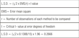 | Table-VI Calculation Of L.S.D.(Least Significant Difference)
|
When the mean optical density of group A-1,A-2 , and A-3was compared it was observed that A-2 i.e. samples in which the post space was prepared seven days after obturation showed minimum leakage (Table I).
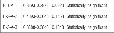
When the optical density of group B-1, B-2 and B-3 was compared it was observed that group B-3 i.e. samples obturated with sectional obturation and post space prepared 14 days after obturation showed minimum leakage (Table II).
If lateral and sectional obturation techniques were compared at the same time interval it was observed that samples obturated with lateral condensation techniques showed less apical leakage (Table III).
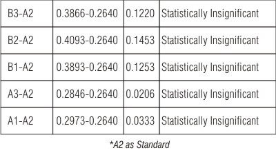
If the readings of all six groups were compared with each other keeping group A-2, which showed minimum mean optical density value, as base group, the inter mean difference was calculated as observed in Table IX. This inter mean difference amongst all the groups was calculated less than least significant difference (L.S.D).
Discussion
Two methods of root canal obturation i.e. lateral condensation and sectional obturation, in teeth to receive post and core preparation, used in this study were designed to closely simulate a clinical situation. Maxillary anterior teeth were selected in this study, since these teeth are more likely to receive post and core preparation as compared to other teeth in the oral cavity because they, being more exposed to extra oral environment, are more prone to fracture in accidents or traumatic injuries.
Lateral condensation technique for obturation of root canals is very commonly employed method. This technique is considered to be effective in sealing the lateral ramifications of the main canals. Such accessory/ lateral canals will remain sealed even after removal of the gutta percha for post space preparation. Therefore this technique was chosen for one of the groups of this study (Group A). Various studies have shown that other obturating techniques yielded no statistically significant difference in apical leakage provided 3-5 mm of gutta percha remained at the apex(10,11,12).For group B ,sectional obturation technique was used to obturate the root canals so as to compare both methods of obturation.
Five millimetres of gutta percha was left intact in the apical region of the root canal as has been advised in a number of studies(10,13,14) Thermal method i.e. hot endodontic plugger, was used to remove the gutta percha from the root canals. Thermal method was preferred over rotary instruments i.e. gates glidden drills and engine driven reamers, because use of rotary instruments is considered to cause greater disturbance of the apical seal which can lead to increased apical leakage(15). Lower leakage pattern of hot instrument method can also be explained b the possibility of a vertical component of the pressure exerted by using the hot plugger for gutta percha removal. This may enhance apical sealing efficacy. In addition, gutta percha has been shown to expand 1-2 % when subjected to heat used in warm instrument technique. However, care must be taken to choose the plugger of correct size for the canal.
Methylene blue 2% was used as the dye for the assessment of marginal integrity at the apex of the specimens. This was preferred over other materials since it is considered a relatively farther penetrating agent with the intensity of penetration being uniform throughout (16).
Four methods are primarily used in dental research to assess degree of marginal leakage,
namely:
1) Linear measurement of dye penetration,
2) Electrochemical analysis,
3) Radio-isotope penetration and
4) Spectrophotometric analysis.
Spectrophotometry as a dye recovery method in dentistry was first described by Douglas and Zakariasen (17). This method minimizes human measurement error and provides determination of volume leakage instead of linear measurements. Spectrophotometry has been shown to be a reliable and effective method for quantitative measurement of apical leakage.
In the present study the results of group A(lateral condensation technique) indicated that there is minimum leakage in subgroup A-2(Mean optical density 0.2640) followed by subgroup A- 3(0.2846). apical leakage was found to be maximum in subgroup A-1(0.2973) (Table I).Reduced leakage in subgroup A-2 can be explained by the fact that a time interval of seven days in the group may be sufficient to harden the cement into the irregularities of the apical third of the root canal leading to closer approximation of the gutta percha to the walls of the root canal. However the difference in the leakage pattern of the three subgroups was minimal and was found to be statistically insignificant (Table VII). The results of the study are in concurrence with other studies concluding that there is no statistically significant difference in apical leakage between teeth that have gutta percha removed immediately after obturation and those in which its removal is delayed by a week for sealer hardening(18).
The results of group B (sectional obturation) indicated that there is minimum leakage in subgroup B-3 (mean optical density 0.3888) followed by subgroup B-1 (0.3893) and subgroup B-2 (0.4992) in that order (Table II). However, the difference in the values of these groups was not statistically significant (Table VIII). The lower leakage observed in group B-3 may be due to hardening of sealer leading to close adaptation of gutta percha to the canal wall in fourteen days which elapsed between obturation of root canal and post preparation. It can therefore be concluded that post preparation at varied time intervals has no definite relationship with the extent of apical leakage. The results of this study are in concurrence with the study of Haddix et al in which no statistically significant difference was found among the lateral condensation , section cone and modified sectional cone techniques in their ability to produce an apical seal(4).
When post preparation in two methods of root canal obturation i.e. lateral condensation and sectional obturation were compared at same time intervals of two, seven and fourteen days (Table III) , it was observed that mean optical density value for A-1 (0.2973) was less than B-1 (0.3993), A-2 (0.2640) less than B-2 (0.4093) and A-3 (0.2846) was less than B-3 (0.3888) i.e. at all time intervals the samples obturated with lateral condensation technique showed less leakage as compared to samples obturated with sectional obturation technique. Less leakage in specimens obturated with lateral condensation technique can be attributed to the physical quality of compressibility of gutta percha. Because of this property the gutta percha can be distorted in the apical third of the root canal and therefore, along with the master cone of the gutta percha additional cones of gutta percha may be used to eliminate voids and truely obturate the root canals in three dimensions. This is not possible in sectional obturation technique. However, the difference in leakage in the two obturating methods at same time intervals was not found to be statistically significant (Table IX).
Mean optical density values of various groups are compared in Table X. Group A-2 i.e. lateral obturation and post space preparation at seven days which showed minimum optical density value , was taken to be standard and all other groups were compared to A-2 and the inter mean difference was calculated. In none of the comparisons the inter mean difference was found to be more than least significant difference (LSD) i.e. 0.2666. It implies that post space preparation in teeth obturated with two different obturating techniques i.e. lateral condensation and sectional obturation at varied time intervals of two, seven and fourteen days did not have any statistically significant different effect on the apical leakage. It is evident that less apical leakage occurs when lateral condensation technique is used for obturating root canals and post space preparation is delayed for seven days. However, the difference in the leakage pattern of various subgroups is statistically insignificant.
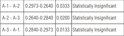 | Table- VII Comparison Of The Lateral Obturation Subgroups At Varied Time Intervals
|
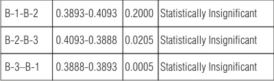 | Table- VIII Comparison Of The Sectional Obturation Subgroups At Varied Time Intervals
|
 | Table-IX The Inter Mean Difference Between Lateral And Sectional Obturation At Same Time Interval
|
 | Table- X The Inter Mean Difference Amongst Various Subgroups Of Group A And Group B
|
References
1. Pappen AF, Bravo M, Gonzalez-Lopez S, Gonzalez-Rodriguez MP. An in vitro study of coronal leakage after intraradicular preparation of cast-dowel space. J Prosthet Dent. 2005 Sep;94(3):214-8.
2. Metzger Z, Abramovitz R, Abramovitz L, Tagger M. Correlation between remaining length of root canal fillings after immediate post space preparation and coronal leakage. J Endod. 2000 Dec;26(12):724-8.
3. Abramovitz I, Lev R, Fuss Z, Metzger Z. The unpredictability of seal after post space preparation: a fluid transport study. J Endod. 2001 Apr;27(4):292-5.
4. Haddix JE, Mattison GD, Shulman CA, Pink FE. Post preparation techniques and their effects on apical seal. J Prosthet Dent. 1990 Nov;64(5):515-9.
5. Hiltner RS, Kulild JC, Weller RN. Effect of mechanical versus thermal removal of guttapercha on the quality of the apical seal following post space preparation. J Endod. 1992 Sep;18(9):451-4
6. Karapanou V, Vera J, Cabrera P, White RR, Goldman M. Effect of immediate and delayed post preparation on apical dye leakage using two different sealers. J Endod. 1996 Nov;22(11):583-5.
7. Solano F, Hartwell G, Appelstein C. Comparison of apical leakage between immediate versus delayed post space preparation using AH Plus sealer. J Endod. 2005 Oct;31(10):752-4.
8. Saunders EM, Saunders WP, Rashid MY. The effect of post space preparation on the apical seal of root fillings using chemically adhesive materials. Int Endod J. 1991 Mar;24(2):51-7.
9. Ewart A, Saunders WP. Investigation into the apical leakage of root-fi- lled teeth prepared for a post crown. Int Endod J. 1990 Sep;23(5):239-44.
10. Camp RL, Todd MJ: The effect of dowel preparation on the apical seal of three common obturation techniques. JPD1983;50(12):664-666.
11. DeNys M, Martens L, Decosten W, Thys M: Evaluation of dowel space preparation on the apical seal using an image processing system. Int Endo J 1989;22(5):240-47.
12. Neagley RL: The effect of dowel preparation on the apical seal of endodontically treated teeth. Oral surg Oral Med Oral Pathol 1969;28(5):739-45.
13. Mattison DG, Delvianis DP: Effect of post preparation on the apical seal. J Pros Dent 1990;64(5):515-19.
14. DyKema RW, Goodacre CJ, Philips RW: Johnston’s modern practice in fixed prosthodontics restoration of mutilated and pulpless teeth. W.B.Saunders and Co. 1986; 4th Ed: pp. 371.
15. Trabert KC, Cooney JP. The endodontically treated tooth. Dent Clin North Am.1984;28(4):934- 35.
16. Matloff IR, Jenson JR, Singer L, Tabibi A: A comparison of methods used in root canal sealability studies. Oral surg 1982;53(2):203-8.
17. Douglas WH, Zakariasen KL: Volumetric assessment of apical leakage utilising a spectrophotometric dye recovery method. J Dent R 1981; 60: 438.
18. Bourgeois R.S, Lemon, R R.: Dowel space preparation and apical leakage. J Endod 1981;7(2): 66-9. |