Introduction
The human mouth provides a suitable habitat for numerous bacterial genera, aerobic as well as anaerobic. The mouth presents a series of different microenvironments, each of which may be colonized by different microorganisms.1 The development of dental plaque has been associated with several environmental and individual factors including diet composition, oral hygiene, fluoride exposure, the quality of saliva, the composition of the oral microflora, and immune factors. Fixed or removable orthodontic appliances also impede the maintenance of oral hygiene, resulting in plaque accumulation2.
Patients undergoing fixed orthodontic appliance treatment have elevated levels of S. Mutans, Candida species and Enterobacteriaceae. Undisturbed supragingival plaque initiates gingival inflammation further leading to gingivitis and gingival hyperplasia3.
There is clear evidence that fixed appliance induce continual accumulation and retention of bacterial plaque 4 and initiates gingival inflammation. There was a study conducted to evaluate the effect of placement of orthodontic bands on microbiological and clinical parameters. The composition of dental plaque determined by dark-field microscopy showed significant shifts in the test sites after banding. Changes consisted of an increase in the percentage of spirochetes, motile rods, filaments, and fusiforms; conversely, a decrease in cocci was noted.5
In a study by Fournier , Payant and Bouclin (1998)6 adherence of streptococcus mutans to the orthodontic brackets , it was proved that saliva coating on bracket surface causes a decreased affinity for streptococcus mutans for all the products . A study for microbiological evaluation of elastomeric chain was done by Casaccia , Gomes , Alviano et. al .(2007)7. in this study the surface of elastomeric chains of different manufacturers were used to verify the presence of pathogenic microorganisms at the moment of unpacking and analyze a possible inhibitory effect of the elastomeric chain when exposed to microorganisms of the oral cavity
It has been proved in various studies that different materials used in fixed mechanotherapy has different rate of microbial growth. Among the different materials used in fixed mechanotherapy, two most commonly used materials for archwire ligation are stainless steel ligature wire and elastomeric rings . The purpose of this study is to compare the microbial colonization associated with two materials used .
Materials And Method
Twenty subjects for this study were selected from Department of Orthodontics and Dentofacial Orthopedics, Subharati Dental College, Meerut.
The following inclusion criteria were used for patient selection:
a. Patients in age group of 11-25years.
b. Initial leveling and alignment completed.
c. Patient on round wires .
d. Patient undergoing fixed orthodontic treatment with brackets on their anterior teeth and bands on their molars.
The following exclusion criteria were used for patient selection:
a. Presence of decalcification of teeth .
b. Presence of anterior composites.
c. Presence of crowding .
The patients were instructed to brush once in the morning before breakfast and once in the evening before bed time. They were instructed to brush a minimum of three minutes to ensure thorough brushing. The patients were asked to thoroughly rinse with water after every meal. The investigation was designed as a split-mouth study. Two commonly used auxiliaries (elastomeric rings and ligature wires) Figure I for tying arch wires were tested. Brackets on the right
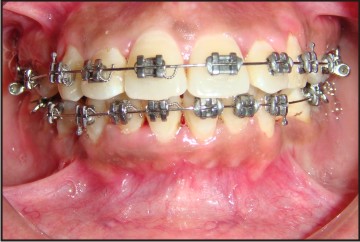 | Figure I - Arch Wire Ligated With Two Different Auxiliaries
 |
side of the dental arch of the patient were ligated with conventional stainless steel ligature wires and brackets on the left side were ligated with elastomeric rings. Microbial records were taken on the first day after thorough oral prophylaxis of patient was done (Table 1), and again after 21 days (Table 2) . On day one, thorough oral prophylaxis of the patient was done and swabs with the help of sterile paper points were taken from the right and the left sides of the upper dental arch .
The arch wire was ligated using two different auxillaries thereafter. On day 21 (Table 2) the conventional stainless steel ligature wire from the bracket of right side second maxillary premolar and the elastomeric ring from the bracket of left side second maxillary premolar was collected and cultivated for aerobic and anaerobic cultures .
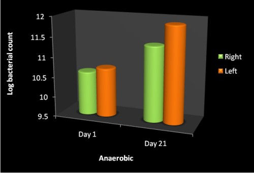
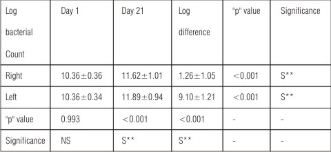 | TABLE - I : Comparison of total anaerobic log bacterial count between Day 1and Day 21
|
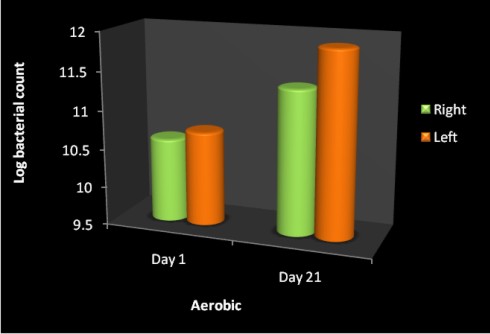
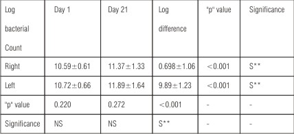 | TABLE - II : Comparison of total aerobic log bacterial count between Day 1 and Day 21
|
Results
The statistical analysis was carried out using SAS 9.2, SPSS15.0, Stata 10.1, MedCalc 9.0.1 and Systat 12.0. All bacterial counts were converted to log bacterial count for ease of statistical calculations. The mean and standard deviations of the bacterial counts values were calculated for both groups. Paired t-test was used to compare the mean anaerobic and aerobic bacterial counts on right and the left side.
Aerobic and the anaerobic bacterial counts were recorded in the study and the data obtained from the right and the left side , day 1 and day 21 , significant difference was found between day 1 and day 21(Graph I and Graph II) observations at 5% level of significance with respect to right and the left sides (Table I and Table II).
Paired "t" test for anaerobic log bacterial count (Table-I) showed significant difference between day 1 and day 21 observations at 5% level of significance with respect to right and the left sides . At day 21 showed significant difference between right and left sides observations at 5% level of significance and no significant difference were found for right and left sides at day1.
Paired "t" test for aerobic log bacterial count (Table-II) showed significant difference between day 1 and day 21 observations at 5% level of significance with respect to right and the left sides . No significant difference were found for right and left sides at day1and day 21.
Discussion
Primary dental care begins at home. Practicing satisfactory oral hygiene, such as adequate tooth brushing, mouth rinsing, and dental flossing, plays a vital role in maintaining healthy teeth, especially in the orthodontic patients8. It is a well known fact that the placement of fixed orthodontic appliances generally hinders good oral hygiene ,and the appliance component can cause alteration in oral micro flora by reducing pH , increasing affinity of bacteria to the metallic surface because of electrostatic reactions, and causing retention areas for microorganisms . Thus they lead to plaque accumulation around the bracket base 2.
However, the contribution of ligation materials to this increase has only been evaluated in few studies. Forsberg et al9 evaluated microbial colonization of 12 patients treated by fixed orthodontic appliances and reported that the lateral incisor attached to the arch wire with an elastomeric ring exhibited a greater number of microorganisms in the plaque than teeth ligated with steel wire. They also reported a significant increase in the number of S. Mutans and lactobacilli in the saliva after the insertion of fixed appliances. They recommended that the use of elastomeric ligation rings should be avoided in patients with inadequate oral hygiene because elastomeric ligation rings will significantly increase microbial accumulation on tooth surfaces adjacent to the brackets, leading to a predisposition for the development of dental caries and gingivitis.
On the other hand, Sukontapatipark et al4 and Turkkahraman 2 evaluated the microbial colonization of 20 patients. Upper second premolar was selected as the donar site, the sample was collected at three different time intervals. They found no significant difference between both materials regarding microbial contamination.
In this study, maxillary second premolars were selected as the donar site for microbial samples because the posterior teeth are more prone to plaque accumulation also access to cleaning is less in posterior region. Bacterial sampling was performed at day 1 when through oral prophylaxis was done and then at day 21, which is equivalent to the average duration between orthodontic appointments. The study was terminated on the third week because longer periods of observation may affect the results as cooperation, motivation for oral hygiene and dietary habits can change.
The result of the current study revealed that the teeth ligated with elastomeric rings exhibited greater number of aerobic as well as anaerobic microorganisms and the difference was found to statistically significant. This result is in accordance with the study of Forsberg et al 9 but is in contrast with the study of Turkkahraman et al 2 who found statistically not significant difference between elastomers and ligatures. A feasible explanation may be due to the difference in the study design i.e. in the present study at the 21 day the elastomeric ring and the ligature was cultured unlike the previous study were the swab from the labial surface of the tooth was taken and in this study anaerobic as well as aerobic culture was done whereas in previous study specifically the growth of streptococcus mutants and lactobacillus was evaluated .
Summary
The present study was done to evaluate the amount of microbial colonization between the two methods of arch wire ligation used i.e. elastomeric rings and stainless steel ligature wires Twenty subjects were selected for the study and were divided into two different groups randomly. All the subjects were brought to the baseline levels after receiving a thorough oral prophylaxis. Aerobic and anaerobic microbial culture was done of the swab collected from labial surface of upper second premolar from right and left sides after receiving through oral prophylaxis. On 21 day the stainless steel ligature wire was collected from second upper premolar of right side and elastomeric ring was collected from the second upper premolar of left side and aerobic and anaerobic microbial culture was carried out .
The mean and standard deviation of both the groups were calculated and statistically analyzed.
The teeth ligated with elastomeric rings exhibited significantly greater number of both the aerobic and anaerobic microorganisms than the teeth ligated with steel ligatures in both the groups.
Conclusion
The following conclusions were drawn from the study:
1. The teeth ligated with elastomeric rings exhibited significantly greater number of both the aerobic and anaerobic microorganisms than the teeth ligated with steel ligatures in both the groups .
2. The anaerobic bacterial count had increased significantly on day 21,on the right as well as the left side.
References
1. Veera L.Sutter. Anaerobes as normal oral flora. JSTOR 1984;6:S62-66.
2. Turkkahraman H, Sayin O, Bozkurt Yesim F, Yetkin Z, Kaya S, Onal S. Archwire ligation technique , microbial colonization and periodontal status in orthodontically treated patients . Angle Orthod 2005;75:231-236
3. Magno Ferreira F A, Enoki Carla, Ito Yoko I, Matsumoto Nakane Aiko M, Faria G, Filho N P. In - vivo evaluation of contamination of super slick elastomeric rings by streptococcus mutans in orthodontic patients . Am J Orthod Dentofac Orthop 2008;133:S104-109.
4. Sukontapatipark W, EI-Agroudi Mohammad A, Selliseth J Nils, Thunold K, Selvig A. Knut. Bacterial colonization associated with fixed orthodontic appliances. A Scanning Electron microscopy study. Eur J Orthod 2001;23:475-484.
5. Huser Coudary M, Baehni C Pieerre, Lang R. Effects of orthodontic bands on microbiological and clinical parameters . Am J Orthod Dentofacial Orthop 1990;97:213-218.
6. Fournier A, Payant L, Bouclin R. Adherence of streptococcus mutans to orthodontic brackets. Am J Orthod Dentofacial Orthop 1998;114:414-417.
7. Casaccia Rembowski G, Gomes C J, Alviano Sales D, Ruellas Oliveira De Carlos A, Anna Sant F E. Microbiological evaluation of elastomeric chains . Angle Orthod 2007;77:890-893
8. Lee JS , Kho Seop H, Lee Woh S, Yang Sik W. Experimental salivary pellicle on the surface of orthodontic materials . Am J Orthod Dentofacial Orthop 2001;119:59-66
9. Forsberg MC, Brattstrom V, Malmberg E, Nord Erik C. Ligature Wires and elastomeric rings: two method of ligation, and their association with microbial colonization of Streptococcus mutans and lactobacilli . Eur J Orthod 1991;13:416-420. |