INTRODUCTION
Authors describe a CAD –CAM1 technique, through the use of advantage of digital photography2 which was based on clinical report. Photographic records aid in identifying esthetics, disharmony and planning for esthetics correction and establishing mutually compatible expectations of dentist and the patient.2 Author consulted text book of Fradeani M3, on esthetic rehabilitation in fixed prosthodontic and esthetic analysis to prosthetic treatment .The case report will cover a patient case report, materials and methods, and final result of the technique for a rotated maxillary right central incisor tooth.
MATERIALS AND METHODS
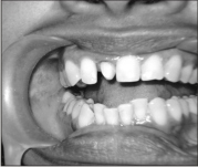
Case report; A 40 years old female patient visited to the deptt. Of Prosthodontics, kothiwal dental college, Moradabad, about three 3 years back. The chief complaint of the patient was mal-aligned right sided maxillary central incisor. She was a professional lady, working as a vice-principal. Examination: extra oral – face profile was convex and intra oral-she had excellent oral health. Her teeth did not have any cavity and periodontal disease. The angle’s class-1 normal occlusion was observed. She also had normal vertical & horizontal overlap. Anterior view showed a right sided mal-aligned (crooked) central incisor (Fig.-1).
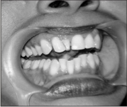 | Fig 1
 |
On Medical examination nothing abnormal was detected. Radiographic examination: A, the IOPA- X Rays disclosed nothing. Bone loss was noticed negligible. Pulpal condition was normal. B, Orthopantogram -NAD (Fig.-2).
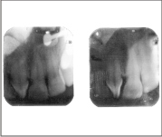 | Fig 2
|
Materials and methods were categorized into non-clinical & clinical. Non clinically, An inter disciplinary approach4, 5, 6 was considered in the form of an orthodontic treatment option, the extraction of crooked incisor, the F P D, R P D, implant, a jacket crown, or the laminate. Authors advocated Computer Assisted Design and Computer Assisted Machining (CAD-CAM) technique for definitive diagnosis and treatment plan as; Non- clinically, Diagnostic casts were made. These casts were mounted on semi- adjustable articulator with centric relation and face bow record transfer. A digital photograph was processed by using a digital camera, having following specifications. Cyber shot DSC-w.30, Sony Japan; 6-mega pixel: sensitivity ISO-1000 (Fig.-3).
 | Fig 3
|
The digital photograph along with dental stone cast was evaluated for a prosthesis. The format and image of the question tooth was adjusted for the space by the application of computer graphic software (CGS) i.e. Photoshop 7.00 adobe system inc. sanjose California .2 It was performed by regaining the space of 2 m.m. from left central incisor and was 1 m.m. from right lateral incisor. An Adjusted final image photograph was obtained from HP2 1315 inkjet printer (Fig.- 4). The resultant two dimensional photographs were shown to the patient for the evaluation of gingival zenith level and future jacket crown (Fig.- 4)
 | Fig 4
|
Clinical procedure; an enameloplasty on left maxillary central incisor and right maxillary lateral incisor was accomplished according to above mentioned dimensions. Reduction of rotated incisor was also performed. Crooked central incisor was reduced in normal manner. A completely reverted7, 8, 9 form of reduced crown was created
(Fig.- 5).
 | Fig 5
|
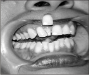
Soft tissue management was carried from double gingival cord procedure9. Final impression was recorded with additional silicon rubber base impression material in custom made special tray. Provisional restoration of self cure acrylic, poly methyl methacrylate was fabricated and cemented with zinc oxide eugenol cement for three weeks. Full veneer crown (FVC) of Zirconia8 via CAD-CAM was fabricated. Finally, it was cemented (Fig.- 6).
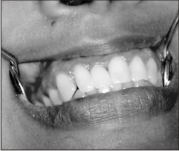 | Fig 6
|
RESULT
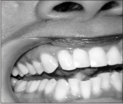
Digital imaging got the advantage of acceptable and deserving natural esthetic treatment modality through two dimensional photographs. These photographs acted like a mediator as immediate treatment option for the patient and an interim aids for clinician and technician as well. This method also changed the treatment option from implant surgery to FDP as an all ceramic crown. Patient was much satisfied and was asymptomatic when reviewed recently.
DISCUSSION
There is no consequence literature available for references regarding the present case. Authors considered that etiologic factors of rotated central incisor could be the injury of child hood in the pre-maxillary region. The Injury displaced and misaligned the developing tooth bud. Adjacent developing teeth buds also misalign the effected tooth. The mid line was slightly shifted; however, Canine position was normal. Because of the shorter space occupied by maxillary arch was adjusted by crowding of mandibular anterior.
Quality has many expressions but indeed targeting perfection is its own reward. Space was occupied by rotated central incisor which was very much less and therefore it gave a distorted gingival zenith position. It affected the future treatment plan options. The main problem was distorted space which was the cause that orthodontic treatment could not be possible (Fig.-1). Implant treatment is not a realistic option. Enameloplasty was done to regain the space for prosthesis. System helps to restore partially mutilated smile and confidence of the patient. However, a special digital photography equipment and computer software system is required. Zirconia, all ceramic jacket crown is the best option amongst the present modalities for anterior prosthesis 8. The final restoration was cemented after three weeks to restore the gingival margin health.
SUMMARY
Present technique could be a wining aid for the diagnosis and treatment options of the discoloured, mutilated, and mal-aligned anterior teeth. Present case report is rare one. Authors, advocated technique can be valuable for the patient and clinician to reduce the psychological questions. Other application of current CAD - CAM technique may be in wider diastema and distorted edentulous spaces as over to an alternative diagnostic teeth set up. It will present a better image to the patient. Consequently, used CAD -CAM may be suggested as an alternative option procedure in the clinics. It absolutely helps to decide the future treatment plan option for a prosthesis.
ACKNOWLEDGEMENTS
I sincerely acknowledge my wife Mrs. Seema singhal and Daughter Ms. Nikhita for their kind cooperation and help.
REFERENCES
1. Glossary of prosthodontics terms. 8th edition. J Prosthet Dent 2005; 94:10-92.
2. Ioli-Ioanna Artopoulou, Peggy j. Wesley, James c. lemon. Digital imaging in fabrication of ocular prostheses. J Prosthet Dent 2006; 95: 327-330.
3. Fradeani M. Esthetic rehabilitation in fixed prosthodontic, esthetic analysis to prosthetic treatment. Chicago quintessence, 2004.
4. T Jan AH, Millwer GD. some esthetics factors in smile, J Prosthet Dent 1984; 51: 24-8.
5. Jain Veena, Gupta R, Duggal R, Prakash H. Restoration of traumatized anterior teeth by inter disciplinary approach – a clinical report. journal of Indian society of pedodontics & preventive dentistry 2005; 23: 197-7.
6. Green HD. A rationale approach top full crown preparation. J Prosthet Dent 1965; 15: 100-5.
7. Malon F.P, Koth L. david. Tylman’s Theory and practice of fixed prosthodontics. 8th Ed; 2000:113-140.
8. Rosensteil F. Stephen, Land F. Martin, Fijimoto Junhei. Contemporary fixed prosthodontics. 4th Ed. St. Lois, Missouri; 2008: 750-800.
9. Fitzig S, Weiss E, Helft M, Metzger Z. A. Gingival guard for crown preparation. J Prosthet Dent 1988; 59:158-60.
|