Introduction:
A laser is a device that emits light (electromagnetic radiation) through a process of optical amplification based on the stimulated emission of photons. The term "laser" was first introduced to the public in Gould's 1959 conference paper "The LASER, Light Amplification by Stimulated Emission of Radiation".1
Lasers have been classified traditionally based on the active medium e.g., gas, liquid, solid state, or semiconductor diode. Whereas, clinically lasers can be classified into two types: soft and hard lasers. The soft tissue lasers available are carbon dioxide lasers, Nd:YAG lasers, argon lasers, Er:YAG lasers, Er,Cr:YSGG lasers and diode lasers. The first laser diode was demonstrated by Robert N. Hall in 1962.2
The diode basically does not interact with dental hard tissues, this makes it an excellent soft tissue surgical laser, indicated for cutting and coagulating gingiva and oral mucosa, and for soft tissue curettage or sulcular debridement.3 A diode laser is a solid-state semiconductor laser that typically uses a combination of Gallium (Ga), Arsenide (Ar), and other elements, such as Aluminium (Al) and Indium (In), to change electrical energy into light energy.4 It is usually operated in contact mode using a flexible fiber optic delivery system, and emits laser in continuous- wave or gated-pulsed modes.3 The power output for dental use is generally around 2 to 10 watts.4 Tissues can respond to laser light in four different ways: scatter, transmit, reflect, and absorb. Absorption is the most desired laser/tissue interaction in dental use which in turn depends on three factors i.e. wavelength, tissue composition and tissue's water content. Laser-tissue interaction is the use of light energy that is absorbed by the tissue to produce a photobiological effect which can be manifested as photodisruption, plasma induced-ablation, photoablation, thermal, and photochemical.5 The mechanisms of diode laser that lead to ablation or decomposition of biological materials are photochemical, thermal or plasma mediated.3
The diode laser exhibits thermal effects using the "hot-tip" effect caused by heat accumulation at the end of the fiber, and produces a relatively thick coagulation layer on the treated surface. Tissue penetration of a diode laser is less while the rate of heat generation is higher than that of the Nd:YAG laser. Since, the diode laser causes minimal damage to the periosteum and bone under the gingiva being treated as well as exhibits the unique property of being able to remove a thin layer of epithelium cleanly, 4 it can be used for a variety of soft tissue procedures without impacting the neighboring tissues. Diode lasers can be used for a multitude of dental procedures which are predominantly soft tissue procedures and include soft tissue surgery, periodontal pocket therapy, peri-implantitis, but can also be used for certain applications involving hard tissue (teeth), i.e. endodontics - root canal disinfection and laser-assisted tooth whitening. The advantages of diode lasers over the other lasers are the smaller size of the units as well as the lower financial costs.4
Regarding advantages of lasers over surgical procedures these include- dry and bloodless surgery, instant sterilization of the surgical site, reduced bacteremia, reduced mechanical trauma, minimal postoperative swelling and scarring and minimal postoperative pain.
These case reports describe few of the various soft tissue procedures that were performed with ezlaseTM 940 nm Diode laser.
Case Reports:
Case 1
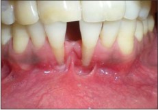
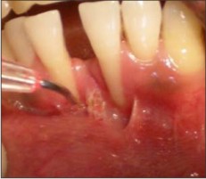
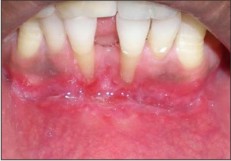
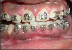
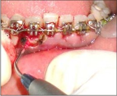
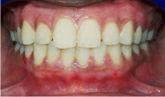
A 21 year old female patient reported to the Department of Periodontics, Babu Banarasi Das College Of Dental Sciences, Lucknow, with the chief complaint of spacing in the upper front tooth region. The patient reported no significant medical history. Presence of a high maxillary frenal attachment was identified as the cause of midline diastema (Fig 1). Therefore the patient had to be treated orthodontically and a frenectomy procedure with diode laser to excise the aberrant frenum was planned. The problem was explained to the patient and a written consent was signed. The frenum was excised with a diode laser in a pulsed mode at a setting of 5 watt, 1 ms pulse interval (Fig 2). The procedure was completed within 10 minutes and caused no discomfort to the patient. The surgical field was clear and dry without bleeding and caused no pain to the patient. No local anaesthesia or suturing was required for the patient. The postoperative area was left to heal by secondary intention (Fig 3). Healing was uneventful and no scarring was seen at 2 weeks (Fig 4).
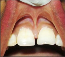 | (fig.1) High Maxillary Labial Frenal Attachment
 |
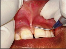 | (fig 2) Frenectomy With Diode Laser
|
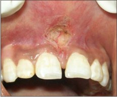 | (fig 3) Postoperative Area
|
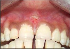 | (fig 4) Healing After One Week
|
Case 2:
A 35 year old male patient reported to the Department of Periodontics, Babu Banarasi Das College Of Dental Sciences, Lucknow,
3 months after the placement of 2 two- stage implants, for the second stage surgery. The implants were placed to replace missing mandibular left second premolar and first molar. Radiograph was taken to ensure complete osseointegration.
Based on the first stage implant placement surgery records of the patient (Fig 5, 6), position of implants were determined and located with the help of trans-gingival probing under topical anesthesia. A written consent was taken from the patient. Diode laser was used at a setting of 2 watt,
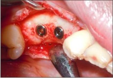 | (fig 5) Preoperative Record (first Stage Implant Surgery- Implant Insertion)
|
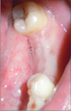 | (fig 6) Preoperative Record (two Weeks After First Stage Implant Surgery)
|
pulsed mode with 0.2 ms pulse interval to excise a circular piece of tissue covering each implant (Fig 7). No anesthesia was required. Cover screws were removed and replaced with healing abutments (Fig 8). Patient reported no discomfort. Gingival margins adapted well and formed a collar around the healing abutments with no shrinkage seen at 1 week.
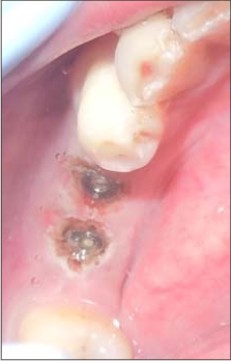 | (fig 7) Second Stage Implant Surgery With Laser
|
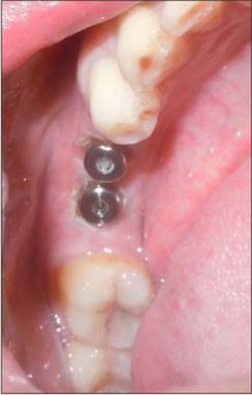 | (fig 8) Healing Abutment Placed
|
Case 3:
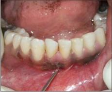
A 20 year old female patient reported to the Department of Periodontics, Babu Banarasi Das College Of Dental Sciences, Lucknow, with the chief complaint of unaesthetic exposure of roots of the lower front teeth, which started 2 years back (Fig 9). A staged approach was planned to treat gingival recession. In the first stage vestibular deepening was done along with frenotomy to increase the width of attached gingiva apical to the recession and to eliminate tension due to mandibular labial frenum on the gingival margins. Recession coverage was planned to be done in the second stage. A written informed consent was taken from the patient. Diode laser at a pulsed mode, 2 watt & 1.0 ms pulse interval was used (Fig 10). The procedure was performed with no patient discomfort or pain. Anesthesia was not required. The healing was uneventful and a gain in the width of attached gingiva could be appreciated after vestibular deepening (Fig 11).
 | (fig 9) Recession In 31, 41 With Shallow Vestibule
|
 | (fig 10) Vestibular Deepening With Laser
|
 | (fig 11) One Week Postoperative
|
Case 4:
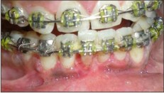
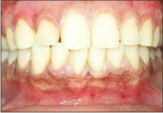
A 19 year old female patient reported to the Department of Periodontics, Babu Banarasi Das College Of Dental Sciences, Lucknow with a chief complaint of gum enlargement in the lower front lower region since 1 month (Fig 12). The patient was undergoing orthodontic treatment since 6 months. The patient reported no medical history and was not under any medication. On intra oral examination gingival enlargement covering half of the crown of the mandibular anterior teeth was observed. Oral prophylaxis was performed, but the enlargement failed to resolve. Therefore a gingivectomy with laser was planned. An informed consent in written was taken from the patient. Diode laser in a pulsed mode at 5 watt and 0.20 ms pulse interval was used (Fig 13). The surgical field was with minimal bleeding. Minimal local anesthesia was injected, and the patient complained of no discomfort or postoperative pain. Healing was uneventful and the gingival contour and size achieved was remarkable (Fig 14)
 | (fig 12) Gingival Enlargement In Mandibular Anterior Region
|
 | (fig 13) Gingivectomy With Laser
|
 | (fig 14) One Week Postoperative
|
Case 5:
A 20 year old male patient reported to the Department of Periodontics, Babu Banarasi Das College Of Dental Sciences, Lucknow with a chief complaint of unaesthetic brownish color of gums (Fig 15). On intra oral examination score-4 gingival pigmentation (Dummett-Gupta Oral Pigmentation Index scoring criteria given by Dummett C.O. in 1964) of the maxillary and mandibular gingiva was observed. The patient was provided with various gingival depigmentation treatment options including conventional scalpel blade, electrocautery and laser. The patient gave consent in written for laser depigmentation. Diode laser in a pulsed mode at 2 watt and 1.00 ms pulse interval was used to treat maxillary gingival hyperpigmentation and 2 weeks later mandibular gingival hyperpigmentation (Fig 16). There was no bleeding, discomfort and the patient was fully satisfied with the results (Fig 17). Even after 3 years of follow-up there were no signs of repigmentation (Fig 18).
 | (fig 15) Gingival Hyperpigmentation
|
 | (fig 16) Laser Depigmentation
|
 | (fig 17) Post Operative Region
|
 | (fig 18) After 3 Years
|
Discussion:
For many intraoral soft tissue surgical procedures, the laser is a viable alternative to the conventional techniques. The commercially available dental instruments have emission wavelengths ranging from 488 nm to 10,600 nm and are all nonionizing radiation. This is to avoid any mutations in the cellular DNA components which ionizing radiations are known to cause.
Different wavelengths have different absorption coefficients based on the varied composition of human tissue. In order to maximize the thermal reaction, there should be a close match between the laser wavelength and the chromophore(s) present in the target tissue. During the thermal ablation as the temperature increases at the surgical site, the soft tissues are subjected to warming (37 to 60°C), protein denaturization, coagulation (> 60°C), welding (70 to 90°C), vaporization (100 to 150°C), and carbonization (> 200°C). The primary chromophores for intraoral soft tissue ablation are hemoglobin, water, and melanin.5, 6 Diode lasers (810-980 nm range) emit laser light in the near infra-red spectrum of the electromagnetic radiation which are highly absorbed in hemoglobin and other pigments. One of the main benefits of using diode lasers is the ability to selectively and precisely interact with diseased tissues. Lasers also allow the clinician to reduce the amount of bacteria and other pathogens in the surgical field, and, in the case of soft-tissue procedures, achieve good hemostasis with the reduced need for sutures. The purported advantages of lasers versus conventional surgery include increased coagulation that yields a dry surgical field and better visualization; the ability to negotiate curvatures and folds within tissue contours; tissue surface sterilization and, therefore, reduction in bacteremia; decreased swelling, edema, and scarring; decreased pain; faster healing response; and increased patient acceptance. When laser cutting is in progress, small blood and lymphatic vessels are sealed due to the generated heat, thereby reducing or eliminating bleeding and edema. Denatured proteins within tissue and plasma are the source of the layer termed "coagulum", which is formed because of laser action and serves to protect the wound from bacterial or frictional action.7 Also the diode laser did not produce any deleterious effect on the root surface. Therefore, diode laser surgery can be performed safely in close proximity to dental hard tissue. All these above mentioned advantages were evidently experienced in the above cases. During procedure, there was no bleeding. Also, postoperatively, no pain was experienced by the patient and no swelling or any other signs of infection were noticed, whereas other alternative procedures have to be accompanied by administration of antibiotics and analgesics to minimize postoperative infection and pain.
In the gingival enlargement case (case 4) reported above, as the patient was wearing fixed orthodontic braces and wires, electro cautery was avoided and surgery with scalpel would have been cumbersome, however treatment with 3mm laser tip provided an indispensable benefit of accessibility and precision.
Using soft tissue lasers is not only beneficial for the patient but for the clinician as well, as in the case of second stage implant surgery with laser, the impression could be taken immediately after implant exposure with little blood contamination in the field due to hemostatic effect of the laser. Also, there is minimal tissue shrinkage after laser surgery, therefore tissue margins remain at the same level after healing as they are immediately after surgery. Different types of dental lasers including diode laser have been used for second stage implant surgery and have demonstrated safety, ease of use, faster recovery and accelerated start of the restorative phase.8
The soft tissue procedures described in the current article validate the advantages associated with diode laser. The risk of eye injury is minimal but must be considered, especially for high-output lasers in the invisible range. Protective goggles, specific for the wavelength, must be used for the patient and the therapist. The future of diode lasers as soft tissue lasers is promising and can be successfully integrated into the everyday dental practice.
Refernces:
1) Gould, R. Gordon. "The LASER, Light Amplification by Stimulated Emission of Radiation". In Franken, P.A. and Sands, R.H. (Eds.). The Ann Arbor Conference on Optical Pumping, the University of Michigan, 15 June through 18 June 1959. pp. 128
2) I A Shcherbakov: Development history of the laser. Physics - Uspekhi 2011; 54: (1): 65 - 71
3) Ameet Mani, Shubhangi Mani, Saumil Shah, Vinayak Thorat: Management of Gingival Hyperpigmentation Using Surgical Blade, Diamond Bur and Diode Laser Therapy: A Case Report. J Oral Laser Applications 2009; 9: 227-232
4) M.L.V. Prabhuji, S.S. Madhupreetha & V. Archana: Treatment of gingival hyperpigmentation for aesthetic purposes using the diode laser. International magazine of laser dentistry 2011; 3: (2): 18-19
5) Niemz MH. Laser-tissue interaction. Fundamentals and applications. 3rd enlarged ed. Berlin, Germany: Springer, 2007:45-46.
6) Goldman L. Chromophores in tissue for laser medicine and laser surgery. Lasers Med Sci 1990; 5(3):289-292.
7) Parker S. Lasers and soft tissue: 'fixed' soft tissue surgery. Br Dent J. 2007 Mar 10; 202(5): 247-253.
8) Martin E. Lasers in dental implantology. Dent Clin North Am 2004: 48: 999-1015, viii.
|