Introduction:
Periradicular surgery is often indicated as a complementary procedure in cases where the conventional endodontic treatment fails. In addition to elimination of pathologic tissues, peri-radicular surgery usually involves apicoectomy, preparation of root end cavity and its filling with a retrograde filling material. During the past decade, newer technologies and materials have been developed for retrograde restorations.. Historically gold foil and amalgam have been used. Subsequently the usage of zinc oxide eugenol, Glass ionomer cement, EBA and Super EBA have been reported in literature.1 Though none of these materials have been proved to provide any osteoinductive and osteoconductive properties.
Mineral trioxide aggregate (MTA) is a powder aggregate containing mineral oxides with a good biological action. MTA when used is able to induce bone regeneration and may facilitate in the regeneration of the periodontal ligament and formation of bone. In addition to these characteristics, MTA presents good physical properties and excellent biocompatibility. As published in literature, it has been hypothesized that MTA may exhibit anti-recurrence properties against the pathological lesions. For a clinical point of view, it has been suggested that MTA can be applied to induce bone formation and inhibit recurrence of the pathology.2
This article reports of a case in which biomaterial such as MTA have been used as a root-end filling material and also to pack the bony defect to induce bone formation.
Case Report:
A 15-year-old female patient was referred to the Department of Conservative Dentistry and Endodontics at Maharishi Markandeshwar College of Dental Sciences and Research, Mullana (Ambala) with the complaint of large swelling in relation to the mandibular central incisors prevailing for over a period of two months. Clinically both the central incisors were tender on percussion and demonstrated Grade-III mobility. In the review of medical history, the patient did not mention any kind of health problems and denied a history of allergies to any pharmaceutical compounds. In dental history review, she stated that she had not suffered any kind of dental trauma or underwent any orthodontic treatment. The patient stated that initially the swelling was small but increased considerably within a span of two months. The radiographic picture presented complete destruction of buccal bone plate and it appeared as if the teeth were "floating" with-in the bone defect. The treatment approach started with the improvement of the periodontal conditions in the surroundings by means of plaque removal, scaling and root planing.
In the next stage, conventional root canal treatment was carried out. Care was taken while performing biomechanical preparation by grasping and supporting the affected teeth between the index finger and the thumb. The root canal was instrumented by hand instruments and obturated according to the technique as explained by Schilder.3 The periradicular surgery was performed immediately after the completion of the endodontic therapy.
The peri-radicular surgery was performed in two phases. The first phase consisted of an apicoectomy with the removal of apical 3mm of root end and preparation of root end cavity by using a bur. Curettage of the lesion was done and haemostasis in the bony cavity was achieved prior to the placement of Mineral Trioxide Aggregate/ MTA (ProRoot MTA, Denstsply, Tulsa). Following all of the aforementioned procedures, MTA was placed in the root end cavity and compacted. Subsequently the entire bony defect was filled up with MTA and the mucoperiosteal flap was repositioned and sutures were placed. To impart immobility and stability to the affected teeth, a composite splint on the facial surface of teeth was placed by achieving support from adjacent teeth. The patient was prescribed appropriate medication including an anti-inflammatory drug and antibiotics. The sutures were removed 7 days post-operatively. Periodic radiographic and clinical examination was carried out which demonstrated considerable healing and the splint was removed after five weeks. Nine months after the peri-radicular surgery, there were no clinical or radiographic signs suggestive of treatment failure, instead the patient's follow-up has demonstrated that the case management has been successful as demonstrated by lesion regression and periodontal repair accompanied with no mobility what so ever.
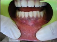 | Pre-operative Clinical
 |
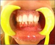 | Post-operative Clinical (After complete healing)
|
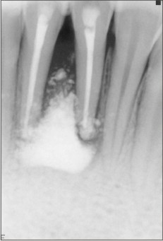
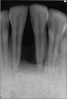 | Pre-operative Radiographic (Teeth appear to be "floating" with-in the bone defect)
|
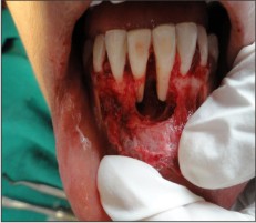 | Large Peri-radicular bone defect
|
 | Postoperative Radiographic (MTA has been filled up in the bone defect)
|
Discussion:
This case report suggests the successful outcome of periradicular surgery after use of MTA as an effective bone replacing material during regenerative tissue procedures. In the case reported in this article MTA was considered as a material of choice because of its characteristics of promoting excellent marginal sealing and stimulating osteoblastic adherence to the retrofilling material surface.4
There were no clinical and radiographical signs suggestive of failure but instead the patients follow up has demonstrated that the case management has been successful as indicated by lesion regression and periodontal repair.
The MTA patent stated that MTA consists of 50-75% (wt) calcium oxide and 15-25% silicon dioxide. These two components together comprise 70-95% of the cement.5 MTA is a powder aggregate containing mineral oxides with a good biological action and may potentiate the regeneration of periodontal ligament and formation of bone.6 Studies have demonstrated that in the presence of MTA, cells grow faster and produce more mineralized gene expression in osteoblasts. Qin H et al have hypothesized that MTA has anti-recurrence properties and inhibit recurrence of such large periradicular defects.2 The most characteristic tissue reaction that MTA exhibits is the presence of connective tissue after the first post operative week.7
The use of biomaterials in this case was necessary because of large destruction of buccal bone plate circum-adjacent to apex of mandibular incisors as chronic lesion associated with affected teeth. It has been reported that MTA promotes the growth of cementum on its surface and reattachment of periodontal ligament so that the contact of extruded MTA with the periapical tissue is not an obstacle to healing.8
Conclusion:
On the basis of the findings of studies addressed in literature review and the clinical and radiographic outcome in this case MTA can be used to induce bone formation and inhibit recurrence of such cases with large periradicular defects.
References:
1. Gatewood RS; Endodontic materials; Dent Clin North Am. 2007 ;51(3):695-712
2. Qin H, Cai J, Fang J, Xu H, Gong Y. Could MTA be a novel medicine on the recurrence therapy for GCTB? ; Med Hypotheses. 2010;74(2):368-369
3. Schilder H. Cleaning and shaping the root canal. Dental Clin North Am. 1974;28:269-296
4. Favieri A, Campos LC, Burity VH, Santa Cecília M, Abad Eda C. Use of biomaterials in periradicular surgery: a case report. J Endod. 2008 ;34(4):490-494
5. Torabinejad M, White DJ. Tooth filling material and use. US Patent Number 5,769,638, 1995
6. Koh ET, McDonald F, Pitt-Ford TR, Torabinejad M. Cellular response to mineral trioxide aggregate. J Endod. 1998;24:543-547
7. Economides N, Pantelidou O, Kokkas A, Tziafas D. Short-term periradicular tissue response to mineral trioxide aggregate (MTA) as root-end filling material. Int Endod J. 2003;36(1):44-8.
8. Johannes Mente, Nathalie Hage, Thorsten Pfefferle, Martin Jean Koch, et al. Mineral Trioxide Aggregate Apical Plugs in Teeth with Open Apical Foramina: A Retrospective Analysis of Treatment Outcome; J Endod; 2009; 35(10);1354-1358 |