Introduction
Knowledge of root canal morphology and possible variations in the anatomy of root canal system is important in the successful management of non-surgical root canal treatment followed by negotiation, cleaning and shaping (Ingle J, 2ND Ed 1976)1
Incomplete instrumentation followed by incorrect obturation is most common cause of failure of as reported by Ingle. Mandibular 2nd pre-molar is typically described as a single rooted tooth with root canal system in text books. There are also numerous case reports and anatomic studies that have reported variations.
Incidence of number of roots and number of root canals reported in anatomic studies varies greatly in literature.2,3
Vertucci in his series of studies conducted on extracted teeth reported 25% incidence of 2nd canal4.
Zilich and Dawson reported 11.7% occurrence of two canals and 0.4% of three canals5,6.
Case of mand. 2nd pre-molar with three canals have been reported by Kaffe I, Kaufmann AY et al 1985, Singh RP , Stamps HF et al 1987, Chan K et al 1992 , Fisher 1992, De Moor RJ 2005 , and Nallapati 2005. Atypical occurance of three canals in 2nd pre-molars with two orifices distal half of furcation area and one orifice on mesial wall of pulp chamber has been reported by De Moor et al7. Various factors like ethnicity , age and gender that can contribute to the differences observed in various anatomic studies have been reported by Troop M , Tronstad I in 19865 and Sert S et al 20048. Apart from morphological studies , frequent case reports have shown unusual root canal variation many of which have been attributed to fusion , germination or concrescence. Present case report is of mandibular 2nd pre-molar with three root canals. The discussion has been based on the significance of such a finding.
Case Report
45 years old male patient reported for the treatment of pain in mandibular right side for last three days. Clinical examination revealed deep disto-occlusal caries with definite pulp involvement. The tooth was tender to percussion. Endodontic treatment was planned for this tooth. Pre-operative radiograph were taken. Careful examination of radiograph showed the presence of two roots and two root canals. Third root canal was not visible because of superimposition.
Treatment was started by giving anesthesia using 2% lidocaine with 1:1,00,000 adrenaline. Endodontic access cavity was prepared through occlusal surface and roof of pulp chamber was removed . Examination showed pulp chamber wide mesio-distally showing two root canals , one mesial and one distal. Continued careful examination to rule out any possibility of additional canal revealed the presence of third root canal in distal root. All three canals were thoroughly instrumented and shaped and obturated with laterally condensed gutta percha using AH plus root canal sealer. Temporary restoration was placed and radiograph was taken. Patient was called after one week for permanent restoration.
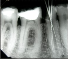 | Fig - 1
 |
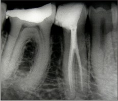 | Fig - 2
|
Discussion
Thorough mechanical cleaning of entire pulp cavity and complete obturation with inert material is main objective of the successful root canal treatment. This objective can not be achieved without thorough knowledge of root canal anatomy , careful interpretation of radiograph and proper modification of access cavity essential for recognition and management of anatomical variations9.
Mandibular 2nd pre-molar is described as a single rooted tooth with single root canal system. The ovoid shaped root in cross section has developmental grooves or depressions on mesial and distal surfaces. The incidence of two separate roots in this tooth is very rare. Often considered an enigma to endodontist, mand. 2nd pre-molar with more than one root and root canals dividing at various levels of the root can generate complex problems.
In the case presented here, two roots with three root canals were identified. Failure to identify 2nd and 3rd root canal might have resulted in incomplete treatment and failure.
Morphological structure in this case is similar to morphological structure studied by Moayedi S , Lata DA and Sanjeev Tyagi et al10.
All these cases involved mand. 2nd pre-molar with two root canals and identical numbers of distribution of root canals. Such similarity should warrant that internal morphology must be identified precisely to identify supplementary root canals or root canal aberrations to achieve successful treatment.
Pulpal floor anatomy and good quality radiograph at different angles is necessary to accurately diagnose the no. of roots and root canals in 2nd pre-molars.
Martinez -Lozano et al11 have suggested 400 mesial angulation of x-ray beam to identify additional canals. Buhrley L J et al 12 and Sempira et al 13 have demonstrated the use of magnification to improve clinical ability to visualize and access canals.
Pulp chamber that appears to be deviated from normal configuration , either triangular or too large in mesio distal plane should be suspected of having three canals (Bellizi and Hartwell- 1981).
Inspite of being difficult to negotiate due to narrowing and curvature , most of the canals can be negotiated and instrumented using current endodontic technique. Failure to recognize the presence of extra root or canals can often lead to acute flare-ups during treatments and subsequent failure.
References
1. Ingle J , Beveridge E : Endodontics 2nd Edition Philadelphia 1976
2. Barre M : The internal anatomy of the teeth with special reference to pulp and its branches ; Dent Cosmos ,1925,67 :581-92
3. Geider P et al : Endodontic anatomy of lower pre-molars Apropos of 669 cases ; J Odontol Conserv 1989 : 11-5
4. Vertucci F J : Root canal anatomy of human permanent teeth Oral Surg Oral Med Oral Pathol 1984; 58 : 589-99
5. Trope M et al : Mandibular pre-molar with more than one root canal in different race groups ; J Endo 1986 , 12;343-345
6. Vertucci F J : Root canal morphology of mandibular pre-molars JADA 97 ;47. 1978
7. De Moor R J et al : Root canal treatment in mandibular 2nd pre-molars with three canals J Endo, 31 ;310-313 : 2005
8. Sert S, Bayirli BS : Evaluation of root canal configuration of mandibular and maxillary permanent teeth by gender in Turkish population J Endo 2004; 30, 391-8
9. Macri E,Zmener O : Five canals in mandibular 2nd pre-molars J Endo 2000; 26, 304-305
10. Sanjeev Tyagi, KP Arjun : Endodontic enigma - Mandibular 2nd pre-molars with three root canals-A case report Peoples Journals of Scientific Research ; vol 1 july 2008 27-29
11. Martinez -lozano et al : of radiographic factors in determining pre-molar root canal system Oral Surg Oral Med Oral Patho Oral Radiol Endod 1999 ; 88 719-22
12. Buhrley L J et al : Effect of magnification on locating the MB2 in maxillary molars J Endo 2002 ; 28, 324-7
13. Sempira et al : Frequency of 2nd MB canal in maxillary molars as determined by the use of an operating microscope- a clinical study J Endo 2000 ; 26, 673-741 |