Introduction:
A sharp bony ridge is a frequent problem among the edentulous patients and commonly occur in the mandible in the edentulous patient. If present should be identified during the initial assessment by palpation of residual edentulous ridges. When it is conventionally loaded, the overlying mucosa is pinched between the denture base and the bone which leads to pain over the ridge.1 Effect in the underlying bony structure of the residual ridge may be the cause of chronic pain under dentures especially during mastication.
Knife edge ridge is formed due to rapid resorption of labial and lingual side of the lower anterior ridge. Gingiva overlying it becomes rolled and soft tissue proliferates leaving hypermobile ridge crest tissue.2
Acc. to Meyer three types of sharp ridges are:2
1) Saw tooth ridge
2) Razor like ridge and
3) Those with discrete spiny projections. However, this classification is academic because all of these type can produce pain under denture
Knife edge ridges are thin, bucccolingually sharp but smooth and like a feather edge they are painful under pressure and this type of ridge seen only in mandible 2
X-ray photographs show a thin ridge with a clearly defined outline, the cancellous bone being covered with a cortical layer3.
Immediate dentures are often an ultimate cause of sharp ridges. Local destruction of the bone by the periodontal disease before tooth extraction, improper surgical procedures of alveolar bone at the time of extraction of teeth, or lack of follow- up and proper correction of changing tissue conditions may be contributing factors.4
A combination of factors contributes to bone resorption, with the amount of resorption and the relative importance of each factors varying with the patients. 4-5
The etiologic agents believed to be of significance include,
1) nutritional inadequacy of the diet
2) endocrine functions
3) tissue resistance to stress
4) traumatic factors (dentures etc)
5) systemic disease and
6) disuse.
The influence of genetic factors appears not to have been investigated.
Inadequate dentures do not necessarily cause residual ridge changes in otherwise healthy individuals.6
Alternative treatments for knife edged ridges:
a) Provision of soft lining-soft lining was not used because of hygiene and maintenance problems associated with these materials.
b) A controlled pressure impression technique would decrease occlusal loading over the affected area and distribute forces more to the primary support areas like buccal self 2
c) Preprosthetic surgery has been widely advocated for dealing with sharp bony ridges. It was not chosen here for the twin disadvantage of surgical trauma to the patient and destruction of potentially stabilizing bone.
d) Differential pressure impression technique. Differential pressure technique was chosen as it enables a conservative preservation of ridge height for stability without overloading the crest of the ridge.
Method
Technique which will distribute loading onto alternative areas over the ridge and relive the mucosa over the sharp bony ridge producing differential pressure, secondary impression of the mandibular arch with a sharp bony ridge
The aim of this technique is to produce loading onto alternative areas (buccal shelf area) and relieve the mucosa over the sharp bony ridge from the load.
The areas that are capable of bearing the load should be preferentially loaded. Those areas that are incapable of load bearing should have their loads reduced1.
Case Report-1
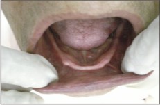
A 60 yr old female patient reported to the Department of Prosthodontics with complains of replacement of her missing teeth. On examination it has been found that patient has very thin (knife edge) ridge present in relation to mandibular arch (fig: 1) thus complete denture has been advised with differential pressure impression technique.
 | Fig - 1
 |
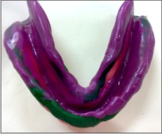
Primary impression was made for both upper and lower arch using impression compound and special tray fabricated on the primary cast. A medium bodied silicone impression was used to make a fully muscle trimmed secondary impression. (fig:2).
 | Fig - 2
|
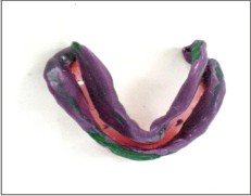
The impression produced displacement of the mucosa over the sharp bony ridge. If it is used to construct the final denture prosthesis, there is a potential for the denture to cause traumatic pain in this region. The area of the impression over the sharp ridge is cut away using a scalpel blade. The tray is perforated over the sharp ridge. (fig:3)
 | Fig - 3
|
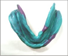
It is important to place numerous large perforations in order to ensure low pressure for the next stage of the impression. Complete impression was made using light bodied impression material (fig: 4). Jaw relation recorded and trying was done.
 | Fig - 4
|
Case No -2
A 56-year old male complained of soreness under his denture. His denture had been made 5 year back. The tissue on the mandibular ridge were hyperplastic, mobile and tender to palpation. Radiograph revealed multiple bony spines protruding from the mandibular ridge crest. The hyperplastic soft tissue and sharp bone were removed surgically. During healing period the lower denture was worn with a soft reline material . new denture were made. The patient reported comfort, retention, and renewed pleasure in eating.
Case No- 3
A 54 year old female patient reported with complained of pain while chewing food. On examination it was found that she had sharp edge ridge in the mandibular arch. She was denture wearer. It was decided that fabricate a new denture for this patient with Differential pressure impression technique. After giving the denture to the patient, she reported one week after and now she don't have any pain on the ridge while chewing the food.
Discussion
The complete denture fabricated by this technique has proved successful for this patient. The successful treatment depended on the accurate diagnosis of the cause of the patient's symptoms.
Bolender and Swenson have reported a successful vestibular extension procedures. This surgical technique might be used for patients who lack adequate ridges after removal of sharp projections to recreate ridge conditions favorable to denture stability and retention.7
The use of the pressure relief areas is a successful form of treatment. However redistribution of the load by the impression technique, as presented here, is likely to create a more controlled loading of the mucosa. Preprosthetic surgery can also be advocated to dealing with the sharp bony ridge. But it is not chosen here because it may lead to trauma to the patient and destruction of the potentially stabilizing bone. Sharp bony ridge cannot bear load but the height of the ridge does provide resistance to horizontal force (stability). Differential pressure impression technique is a conservative preservation of the ridge height for stability without overloading the underling mucosa1.Thus selection of the appropriate impression technique is essential for the success of our prosthesis.
References
1. T P Hyde" a case report ;differiential pressure impression for complete denture : Eur J Prosthodont Restro Dent, 2003 mar; 11 (1) 5-8.
2. Sheldom Winkler , essential of complete denture prosthodontics. A.I.T.B.S. Publishers and distributors 2nd edition 2000.
3. H.R.B. Fenn, K.P. Liddelow, A.P. Gimson, clinical dental prosthetics, CBS Publisher 1st edition1986.
4. Roger A. Meyer; Management of denture patients with sharp residual ridges. j pros. Dent; may- june, 1966; 16 (3) 431-37.
5. Clinton F. Sobolik; alveolar bone resorption. j. pros dent. 1960 july- august ; 10 (4) 612-19
6. Atwood, D. A. ; some clinical factors related to rate of resorption of residual ridges. j. pros. Dent.1962; 12: 441-50.
7. Bolender, C. L., Swenson, R. D: cephalometric evaluation of a labial vestibular extension procedure. j pros. Dent. 1963; 13: 416-31 |