Introduction
"A new eruptive fever with stomatitis and opthalmia" was described as a severe variant of erythema multiforme & was termed by Steven and Johnson in 1922. By the 1940's it was commonly called as "Steven Johnson's syndrome (SJS)". The concept of the spectrum of erythema multiforme has been widely accepted since that time1. SJS is an acute, self-limited disease, Presenting as severe mucosal erosions with widespread erythematous, cutaneous macules or atypical targets2.
Although SJS is rare with an incidence of 0.05 to 2 persons per 1 million populations per year, it has significant impact on the public health in view of its high morbidity and mortality3. Majority of cases are drug- induced4.
SJS is one such disease which could manifest with extensive oral and peri-oral involvement, where oral physicians come into picture. Hence we are presenting a case of SJS, which has been successfully treated.
Case Report
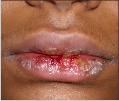
16 year old female came to our Department with a complaint of painful ulcers in the mouth & bleeding from lips since a day. She presented with fever a week back for which she was prescribed antibiotics & analgesics after which she developed ulcers in the mouth & bleeding from lips, associated with pain which was sudden in onset, burning type, continuous, localized, and severe in intensity, aggravated on touching, speaking, eating food & there was no relieving factor. She also had burning micturation & watering of both eyes since 4 days. Her past medical history revealed that she was suffering with fever a week back & was on tab. Ciprofloxacin 500mg tid for 5days & tab Paracetamol 500mg + Diclofenac sodium 50mg tid for 3days for the same. Her past dental & surgical histories were non contributory. Patient had elevated temperature of 100F. Bilateral submandibular lymph nodes were palpable, tender, mobile, firm in consistency. Extra orally there was limited mouth opening & erythematous crusted areas on both upper & lower lip extending upto the vermilion border with fresh bleeding spots, surface appeared rough & scaly, on palpation the periphery of the lesion was hard & tender (figure 1).
 | Figure 1 - Erythematous Crusted Areas On Both Upper & Lower Lip Extending Upto The Vermilion Border With Fresh Bleeding Spots
 |
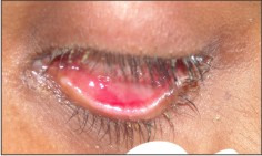
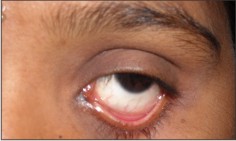
Bleeding was evident on touching with peeling up of overlying skin of lips. There was solitary ulcer seen in conjunctiva bilaterally, associated with watering of eyes & pus discharge (figure 2).
 | Figure 2 - Solitary Large Ulcer On The Conjunctiva Bilaterally
|
The vaginal lesion was confirmed with examination in department of Veenerology. Bilateral solitary submandibular lymph nodes were palpable, tender, mobile, firm & measured 1x1 cm. Intra oral examination revealed clusters of ulcers on lower labial mucosa, right & left buccal mucosa (figure 3) & right lateral border of tongue (figure 4) with fresh bleeding spots, each ulcer measured about 1×0.5 cm, surrounded with erythematous halo. Floor of ulcers revealed a whitish slough. Slight provocation initiated bleeding.
& right lateral border of tongue (figure 4) with fresh bleeding spots, each ulcer measured about 1×0.5 cm, surrounded with erythematous halo. Floor of ulcers revealed a whitish slough. Slight provocation initiated bleeding.
Based on this our clinical diagnosis was Stevens Johnson Syndrome as the lesion noticed in eyes & genitals. Differential diagnosis thought were phemphigus vulgaris & stomatitis medicamentosa. We could subject the patient to only the hematologic investigation as the lesion being acute; the patient was under severe discomfort. Her complete blood picture revealed raised ESR - 50mm/1st hr & total leucocyte count was 12000 cells/mm3. Rest other findings were within normal range.
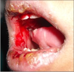 | Figure 3 - Fresh Bleeding Spot With Ulcers On Right Buccal Mucosa
|
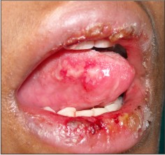 | Figure 4 - Clusters Of Ulcers On Right Lateral Border Of Tongue
|
We treated her with systemic steroids, tab prednisolone 10mg qid for 7 days, which was gradually tapered to 10mg tid for 7days, 10mg bid for 5days & 10mg once daily for 5days respectively, benzydyamine hydrochloride 0.15% oral rinse for oral ulcers. Gention violet application for lip lesions. Clotrimazole cream 1% for vaginal lesion & ofloxacin eye drops 0.3% for eye lesion. Liquid & soft diet was advised. All the lesions healed within 15 days (figure 5); there was absence of burning micturation & lacrimation (figure 6).
 | Figure 5 - 15 Days After The Treatment Shows Complete Healing Of Ulcers
|
 | Figure 6 - Eye Lesions Healed
|
Discussion
Stevens-Johnson syndrome is a severe, episodic mucocutaneous intolerance reaction described by Hebra5 in 1866 and Albert Mason Stevens and Frank Chambliss Johnson in 1922. Erythema multiforme (EM) and Stevens-Johnson syndrome are part of a clinical spectrum6.
SJS is said to be associated with drugs. More than 100 different medications have been implicated. The syndrome usually begins within 1-14 days of ingestion of the offending agent7. In our case the interval was one week. SJS and TEN are considered T cell mediated disorders in which activation of CD8 T lymphocytes lead to destruction and apoptosis of keratinocytes8. Drugs can activate T cells by acting as a hapten, as a prohapten or by direct pharmacologic interaction among the drug, Major Histocompatibility Complex (MHC) molecule and a T cell receptor. It is postulated that drugs can bind with the MHC and T cell receptor causing activation of T cells contributing to SJS. Yap et al3 determined that the drugs especially anticonvulsants and allopurinol were the major causes of SJS. Jean-Claude Roujeau, et al9 in 1995 conducted a study on medication use & risk of SJS & TEN, according to whom the commonest drugs causing SJS included most classes of antibiotics, including cephalosporins, quinolones, aminopenicillins, tetracyclines, and imidazole antifungal agents. A recent study by Gokhan Okan, et al7 in 2008 reported a case of ciprofloxacin induced SJS. This holds well in our case where the patient was on ciprofloxacin for a week. Kristina E, et al10 stated that among the available NSAIDs, oxicam derivatives have the greatest association with SJS & the risk of SJS or TEN in patients receiving NSAIDs is extremely low; whereas older patients, women, and patients within the first month of treatment initiation have the greatest risk. Mainly drug- induced SJS-TEN Overlap and TEN have high mortality. The reported mortality rate ranges from 5% to 40% 11, 12. The syndrome may be diagnosed on clinical criteria. Signs and symptoms consist of a non-specific prodrome of malaise, fever, headache, sore throat, cough, chest pain, vomiting, diarrhea or myalgias6.
Systemic corticosteroids has unproven benefit in early cases of SJS and TEN and deleterious in the advanced forms13. Other treatment includes cyclosporine and intravenous immunoglobulin but new treatments, such as amniotic membrane support for ocular damage, may need to be considered14. To conclude drug induced SJS is more common & is considered to have high mortality rate, herewith we presented a case of drug induced SJS which was treated successfully with systemic steroids.
References
1. Fitzpatrick's dermatology in general medicine, Editors: Irwin m. Freeberg, Arthur Z. Eisen, Klans Wolff, K. Frank Austin, Lowell A. Goldsmith, Stephen I. Katz, 6th edition, Mc Graw Hill, 2003, page: 543-557
2. JP Fagot, et al. Nevirapine and the risk of Stevens-Johnson syndrome or toxic epidermal necrolysis. AIDS 2001, 15:1843-1848
3. FBB Yap, M Wahiduzzaman, & M Pubalan. Stevens - Johnson syndrome (SJS) and Toxic Epidermal Necrolysis (TEN) In Sarawak: A Four Years' Review. Egyptian Dermatology Online Journal 2008; 4(1): 1-13
4. MD Kamaliah, D Zainal, N Mokhtar, N Nazmi, Erythema multiforme, Stevens Johnson syndrome and toxic epidermal necrolysis in north eastern Malaysia, Int J Dermatol. 37: 520- 3, 1998.
5. AM Stevens, FC Johnson. A new eruptive fever associated with stomatitis and ophthalmia. Amer J Dis Child 1922; 24:526-533
6. M Dinerman. Stevens-Johnson Syndrome with Mycoplasma pneumoniae and Enterovirus. International Pediatrics 2004; 19(4): 237-239
7. Gokhan Okan, et al. Vanishing bile duct and Stevens-Johnson syndrome associated with ciprofloxacin treated with tacrolimus. World J Gastroenterol 2008 August 7; 14(29): 4697-4700
8. J Kehren, C Desvignes, M Krasteva et al., Cytotoxicity is mandatory for CD8 (+) T cell-mediated contact hypersensitivity, J Exp Med. 189: 779- 86, 1999
9. Jean-Claude Roujeau, et al. Medication Use and the Risk of Stevens-Johnson Syndrome or Toxic Epidermal Necrolysis. The new England journal of medicine 1995; 333(14): 1600-1608
10. E Kristina, Ward, R Archambault and L Tracey. Mersfelder. Severe adverse skin reactions to nonsteroidal antiinflammatory drugs: A review of the literature. American Journal of Health-System Pharmacy 2010; 67(3): 206-213
11. A Auquier-Dunant, M Mockenhaupt, L Naldi, O Correia & W Schroder. Correlations between clinical patterns and causes of erythema multiforme majus, Stevens-Johnson syndrome and toxic epidermal necrolysis, Arch Dermatol. 2002; 138: 1019- 24
12. PD Ghislain & JC Roujeau, Treatment of severe drug reactions: Stevens- Johnson syndrome, toxic epidermal necrolysis and hypersensitivity syndrome, Dermatol Online J 2002; 8(1): 5.
13. RA Stuart, PJ Gow, N Bellamy, J Campbell & R Grigor. A survey of current prescribing practices of anti-inflammatory and uratelowering drugs in gouty arthritis, NZ Med J 1991; 104: 115-7.
14. S Knowles & NH Shear. Clinical risk management of Stevens-Johnson syndrome/toxic epidermal necrolysis spectrum. Dermatol Ther 2009; 22(5):441-51. |