Introduction
In today's world of globalization, most of our dental patients are aware and conscious of their looks. They may lack the technician knowledge of fabrication of a prosthesis or restoration but can very well make out the difference between an artificial looking and a natural looking restoration /crown which does match in size, shape and color with the adjacent natural teeth. Hence it is of utmost importance for the dentist to select the shade of the artificial teeth to be an exact replica of their natural teeth. Hence shade selection becomes a very important criterion for success of the dentist in satisfying the patient with esthetically superior restorations.
Most of our dental fraternity uses the traditional method for selecting shade which is based on the experience and the judgment of the trained human eye of the operator. But not all dental personnel are lucky to have a perfect color vision.
Dentists having defective color vision may be unaware of their defect or may have problems in perceiving color as normal vision dentists do. "Color blindness" is a misnomer as only a small percentage of people are unable to see any color.
Hence the term can be replaced by color vision defect. Color vision depends on the absorption of light by visual pigments contained within specialized cells in the eye called photoreceptors. Cones are responsible for color vision. The human retina has cone cells which see mainly red, green and blue. Other colors are interpreted as mixtures of these. People who are "color vision defective" tend to be missing some of the color-sensitive cones, so these colors will appear darker.
Color defective vision is either inherited or acquired. Defect can be acquired as a result of eye diseases or normal aging or as a side effect of some medications. In acquired defects, other parts of the eye besides cones and cone pigments may be affected. There are three groups of inherited color vision defects:
1.Monochromacy: Rod monochromats, or complete achromats are truly "color blind" since they cannot distinguish any hues (e.g., blue, green, yellow and red). Different degrees of lightness can be seen by them. The world appears to be shades of gray, black and white
2.Dichromacy: less severe than monochromacy, can distinguish some colors. Dichromacy is divided : protanopia, deuteranopia and tritanopia. Protanopia and deuteranopia are red-green defects. Persons with red-green defects cannot distinguish between red, greens and yellow but can discriminate between blue and yellow. Protanopes often can identify red and green correctly because green looks lighter to them than red. Hereditary tritanopia is rare, a blue-yellow defect. Persons with blue-yellow defects cannot see the difference between blue and yellow but can distinguish between red and green.
3.Anomalous Trichromatism : sensitivity to all three hues, with abnormality in retinal cones affecting one of primary pigments. A need was felt to atleast screen the dental personnel to make them aware of the possible color defect they may be having which may lead to wrong shade matching and hence repeating their restorations. This study could help them is modifying their method of shade matching.
Materials and Methods
A sample size of 400 was randomly selected for the screening study comprising of dental students, dental teaching faculty and dental technicians/ auxillaries. 200 among the sample size were male and 200 female with age ranges from 17 to 35 years. Ishihara color blindness test(numbers made up of color dots) ( Fig.1) was conducted to screen the color deficient individuals in the same room and in the same light source. Ten templates were prepared comprising of six circles. Each circle comprised of a number made up of colored dots as shown in the figure. Each study sample was asked to read the number from the colored circles. All other variables that would affect the study were kept constant for all the individuals.
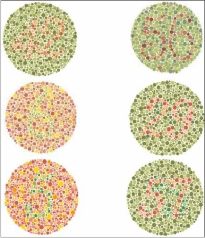 | Fig.1 Ishiara color blindness test. Sample template: identify the numbers in the above colored circles.
 |
Eg:
1) source of light and the room for study was kept the same for all the individuals
2) Eye fatigueeach individual was given four seconds to identify the number
3) Test was carried out at the same time of the day.
Normal vision personnel could decipher the numbers.Individuals who were not able to decipher the numbers or read the numbers incorrectly were marked as suspected color vision defects and the test was repeated for them. The data was analyzed and conclusions drawn.
Results
10 individuals were suspected of color vision defect. All the individuals were male (5% of the male sample study). None of the females were found to be color blind. All the individuals could differentiate between blue and red spots but all the color deficient subjects couldn't differentiate between green and red spots.
Discussion
Color blindness is one of the common genetic disorders observed. It is a sex linked recessive trait. The genes are located on the X chromosome within the Xq28 band. If a man is a carrier of a defective X-chromosome he will suffer from color blindness. On women the not defective chromosome is in charge and therefore she is not colorblind but a carrier for color blindness. In our study it was found that 5% of the males were color blind while 0% of the females were color blind. This result was almost comparable to many studies done on the same ground. In the study conducted by Al Aqtum, it was found that 0.33% females were color blind: one of them showed protanomalia; 1 protanopia; and 2 deutranopia. In males - 8.72% were color blind: 4 showed protanopia, 8 deuteranomalia and 4 deutanopia. 1 In the another study, it was found that 9.3% of men and none of the women were color blind.2 The prevalence of color defective vision in male dentists was found to be 8.2% by Mc Maugh,3 9.9% by Moser et al4 and 14% by Barna et al.5 Previous studies have shown that color defective personnel were found to make significant errors in hue and chroma selection than normal vision people.6 Observers were more sensitive and critical of crowns where color differed in redness as opposed to crowns whose color differed to the same extent in yellowness.7
There are many tests for color blindness eg. Pseudoisochromatic plates like Ishihara test and Dvorine, Bostrom, AO HRR, Farnsworth-Munsell 100 Hue Test which gives a person many colored caps with slight variations of colors, and asks him to sort colors that are very close together.
However, it is slow and expensive to administer by a specialist, and is not common. Anomaloscope is a better test requiring a specialist which gives exact result of red and green problems. In our test, Ishihara plates were used to screen the individuals. It is not a confirmatory test and it was only used for screening defective vision people who were later advised to refer a specialist for confirmatory diagnosis. The personnel suspected with a color defect can be counseled on other alternate options of shade matching instead of the traditional trained human eye method. Electronic shade matching devices like colorimeters, spectrophotometers and digital color analyzers are available. Colorimeters use photodiode filters to control light reaching the specimen. The light reflected from the specimen is then measured by a detector. Colorimeters are easier to use and are less expensive than spectrophotometers. However, repeatability may be poor due to aging of filters, and object metamerism can be a challenge to their accuracy.8,9Of all devices, a spectrophotometer is the most accurate for absolute color measurement. These instruments have a longer working life than colorimeters and are unaffected by object metamerism.8-11
There are many self contained systems of shade matching available: They include the VITA Easyshade by Vident, Spectroshade by Posey Dental Technology, Shadeye NCC by Shofu, Shadescan by CYNOVAD, and Shadevision by XRite. Color measurement is determined either by a colorimeter (Shadye NCC, and Shadevision), spectrophotometer (Vita Easy Shade and Spectroshade), or a proprietary system called "artificial vision" (Shadescan) ShadeVision (X-Rite, Inc, Grand Rapids, Mich), SpectroShade Micro (MHT SpA, Verona, Italy) and Easyshade (VITA Zahnfabrik). Spectrophotometers generally can provide more systematic and precise measurements than colorimeters because of their ability to measure the amount of light reflected from objects over a full spectral wavelength.
Two-dimensional image capture also provides a visual image of the target tooth. It can also be suggested to have an assistant trained in color matching to the operator, who has color defective vision.
Summary and Conclusions:
Males (5%) show color defective vision more than females( 0%) due to its genetic predisposition. Dental students and personnel were screened for color defective vision and referred to the ophthalmologist for more accurate investigations. Alternate means of shade selection/ matching be advised for color defective personnel.Defective color vision students and personnel were advised to take assistance in appointments of shade selection / matching.
References
1. Al-Aqtum MT, Al - Qawasinch MH. Prevalence of color blindness in young Jordanians. Ophthalmologia 2001;215(1);39-42.
2. Wasson W, Schuman N. Color vision and dentistry. Quintessence Intl. 1992;23:349-53
3. McMaugh DR. A comparative analysis of the color matching ability of dentists,dental students, and ceramic technicians. Aust Dent J 1977;22: 165-167.
4. Moser JB, Wozniak WT, Naleway CA, et al. Color vision in dentistry: A survey.JADA 1985;110:509-510.
5. Barna GJ, Taylor JW, King GE, Pelleu GB. The influence of selected light intensities on color perception within the color range of natural teeth. J Prosthet Dent 1981;46:450-453.
6. Davison SP, Myslinski NR. Shade selection by color vision defective dental personnel. J Prosthet Dent 1990;63:97-101
7. Douglas RD, Brewer JD. Acceptability of shade differences in metal ceramic crowns. J Prosthet Dent. 1998;79:254-260.
8. Paravina RD, Powers JM. Esthetic color training in dentistry. St. Louis: Mosby; 2004.,17-28, 169-170
9. CIE (Commission Internationale de l'Eclairage). Colorimetry, official recommendations of the International Commission on Illumination,: Colorimetry, 3rd ed ,Publication CIE No.15:2004 |