|
|
|
| Treatment Of Gingival Hyperpigmentation By Scalpel Surgery And Electrosurgery : A Split Mouth Design |
Shankar T. Gokhale 1 , Vatsala V. 2 , Rohit Gupta 3 , Ira Gupta 4
1 BDS, MDS, Professor, Department of Periodontics - Rama Dental College & Hospital, Kanpur, (U. P.)
2 BDS, Post Graduate Student, Department of Pedodontics & Preventive Dentistry - Rama Dental College & Hospital, Kanpur, (U. P.)
3 BDS, MDS, Reader, Department of Periodontics - Rama Dental College & Hospital, Kanpur, (U. P.)
4 BDS, MDS, Reader, Department of Periodontics - Rama Dental College & Hospital, Kanpur, (U. P.)
|
| Address For Correspondence |
Dr. SHANKAR T. GOKHALE,
Professor, Department of Periodontics,
Rama Dental College & Research Centre,
A/1-8, Lakhanpur, Kanpur-208024, (U.P.)
Phone: +919794200404
E-mail: dr_shankartg@yahoo.co.in |
| Abstract |
| Gingival hyperpigmentation is mostly caused by the physiologic deposition of melanin by melanocytes. In "Gummy Smile" Patients, melanin gingival hyperpigmentation causes an esthetic problem and may cause physiologic disturbances. Methods to remove gingival hyperpigmentation vary but here in this present case report a split mouth design was used to compare scalpel surgery and electrosurgery procedures. |
|
| Keywords |
| Depigmentation, Gingiva, Scalpel surgery and Electrosurgery Procedures |
|
| Full Text |
Introduction:
The color of the attached and marginal gingival is generally described as pink. It is determined by several factors, including the number and size of blood vessels, epithelial thickness, quantity of keratinisation and pigment within the epithelium.
Gingival hyperpigmentation occurs as a diffuse, deep-purplish discoloration or as irregularly shaped brown and light brown patches. The distribution of oral pigmentation in black individuals is as follows: gingival = 60%; hard palate = 61%; mucous membranes = 22% and tongue = 15%. [1]
Melanin hyperpigmentation of gingiva usually does not present as a medical problem, but many patients may consider their black gums to be unaesthetic. This problem is aggravated in patients with a "gummy smile" or excessive gingival display while smiling. Gingival depigmentation is a periodontal plastic surgical procedure whereby the gingival hyperpigmentation is removed or reduced by various techniques. The foremost indication for depigmentation therapy is the demand by a person for improved esthetics.
Demand for cosmetic therapy of gingival hyperpigmentation is common. Various methods as gingivectomy,[2] gingivectomy with free gingival autografts,[3] acellular dermal matrix allografts,[4] electrosurgery,[5] cryosurgery,[6] abrasion with diamond bur,[7] and various types of lasers[8] have been used for cosmetic therapy of gingival melanin depigmentation.
The present case report compares two techniques i.e., a simple and effective surgical depigmentation using scalpel on one side and electrosurgery on contralateral side in a split mouth design.
Case Report
A 20 year old female patient had a chief complaint of having unesthetic, diffuse, dark-brown to black gingival discoloration in the labial aspect of the maxilla and mandible (Fig. 1).
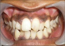 | Figure 1. Pre-operative picture of 20 year old female complaining of black coloured gums.
 |
The depigmentation treatment procedure was carried out in the department of Periodontics, Rama Dental College Hospital, Research Centre, Kanpur, Uttar Pradesh, India.
A scalpel surgery with bur abrasion on one side and electrosurgery on contralateral side was planned to perform the depigmentation. The entire procedure was explained to the patient and written consent was obtained. A complete medical, family history and blood investigations were carried out to rule out any contraindication for surgery. Local anesthesia was infiltrated in the maxillary anterior region from premolar to premolar ( Lignocaine with adrenaline in the ratio 1:100000 by weight).
Electrosurgery technique was performed with the needle electrode, supplemented by the small, ovoid loop or diamond-shaped electrodes for festooning. A blended cutting & coagulating (fully rectified) current was used. In all the steps the electrode was activated and moved in a concise "shaving" motion. Extreme care was exercised to avoid contacting the tooth surface. For hemostasis, the ball electrode was used (Fig. 2).
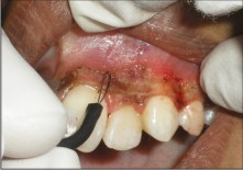 | Figure 2. Electrosurgery being used to remove the pigment layer in maxillary anterior gingiva (21 to 24).
|
On contralateral side a Bard Parker handle with a No. 15 blade was used to remove the pigmented layer. Pressure was applied with sterile gauze soaked in local anesthetic agent to control hemorrhage during the procedure. After removing the entire pigmented epithelium along with a thin layer of connective tissue with scalpel, abrasion with diamond bur was done to get the physiological contour of the gingival, the exposed surface was irrigated with saline. While using the bur minimal pressure was applied with feather light brushing strokes and without holding bur in one place. Care was taken to see that all remnants of the pigment layer was removed (Fig. 3).
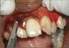 | Figure 3. No. 15 blade being used to remove the pigment layer in maxillary anterior gingival (11 to 14)
|
Post-surgical instructions were given to the patient along with antibiotics (Amoxicillin 500 mg, thrice daily for five days) and Antiinflammatory Analgesics ( Ibuprofen and Paracetamol thrice daily for three days). The patient was advised to 0.2% chlorhexidine gluconate mouth wash 12th hourly for one week.
The patient was reviewed at the end of one week. The healing process was proceeding normally and it was quite uneventful on scalpel surgical area than compared to electrosurgical area and patient did not report any discomfort. The patient was asked to continue the chlorhexidine mouth wash for another week. At the end of one month, re-epithelization was complete and healing was found to be satisfactory. Patient had no complaints of post-operative pain or sensitivity. The gingiva appeared healthy and no repigmentation was observed (Fig. 4).
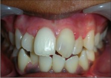 | Figure 4. Six months post-operative picture showing healthy gingiva with no recurrence.
|
Discussion
Oral pigmentation occurs in all races of man. There are no significant differences in oral pigmentation between males and females. The intensity and distribution of pigmentation of the oral mucosa is variable, not only between races, but also between different areas of the same mouth.
Melanin pigmentation was is frequently caused by melanin deposition by active melanocytes located mainly in the basal layer of the oral epithelium. Pigmentation can be removed for esthetic reasons. Different treatment modalities have been used for this purpose.[4] The selection of a technique for depigmentation of the gingiva should be based on clinical experience, patient's affordability and individual preferences.
Scalpel surgical technique is highly recommended in consideration of the equipment constraints that may not be frequently available in clinics. It is known that the healing period for scalpel wounds is faster than other techniques.[9]
Electrosurgery requires more expertise than scalpel surgery. Prolonged or repeated application of current to tissue induces heat accumulation and undesired tissue destruction. Contact with periosteum or alveolar bone and vital teeth should be avoided.[10]
Post surgical repigmentation of gingiva has been previously reported. Repigmentation is described as spontaneous and has been attributed to the activity and migration of melanocytic cells from surrounding areas. In the present case no incidence of repigmentation was observed at the end of one month. The case is being followed up to estimate further the extent and rate of pigmentation.
Conclusion
The depigmentation procedure was successful and the patient was satisfied with the result. Hence we conclude that depigmentation of hyperpigmented gingiva by scalpel surgery with bur abrasion is simple, easy to perform, cost effective and above it all it causes less discomfort. On the otherhand electrosurgery procedure provided blood free working area and achieving contouring and festooning was easy with various electrodes. As far as healing was concerned, it was relatively better with scalpel surgery compared to electrosurgery at the end of one week but no difference was found at the end of one month.
References
1. Dummett CO. Physiologic pigmentation of the oral and cutaneous tissues in the Negro. J Dent Res 1946; 25: 421-432.
2. Bergamaschi O, Kon S, Doine AI and Ruben MP. Melanin repigmentation after gingivectomy: A 5 - year clinical and transmission electron microscopic study in humans. International journal of Periodontics and Restorative Dentistry 1993; 13(1): 85-92.
3. Tamizi M, Taheri M. Treatment of severe physiologic gingival pigmentation with free gingival autograft. Quintessence Int 1996; 27: 555-558.
4. Pontes AE, Pontes CC, Sovza SL, Novaes AB (Jr.), Grisi MF, Taba M(Jr.). Evaluation of the of efficacy of the acellular dermal matrix allograft with partial thickness flap in the elimination of gingival melanin pigmentation. A comparative clinical study with 12 months of follow - up. Journal of Esthetic and Restorative Dentistry 2006; 18(3): 135-143.
5. Gnanaesekhar JD, Al - Duwairi YS. Electrosurgey in dentistry. Quintessence International (Berlin) 1998; 29(10): 649-654.
6. Yeh CJ. Cryosugical management of melanin pigmented gingiva. Oral Surg Oral Medicine Oral pathology Oral Radiology Endodontology 1998; 86(6): 660-663.
7. Bishop K. Treatment of unslightly oral pigmentation: A case report. Dental update 1994; 21(6): 236-237.
8. Stabholz A, Zeltser R, Sela M, Peretz B, Moshonov J, Ziskind D. The use of lasers in dentistry: principles of operation and clinical applications. Compendium of continuing Education in Dentistry 2003; 24(12): 935-948.
9. Almas K and Sadiq W. Surgical treatment of melanin pigmented gingiva: An esthetic approach. Indian journal of Dental Research 2002; 13(2): 70-73.
10. Ozbayrak S, Dumla A, Ercalik - Yalcinkaya S. Treatment of melanin-pigmented gingiva and oral mucosa by CO2 laser. Oral Surgery Oral Medicine Oral Pathology Oral Radiology and Endodontics 2000; 90(1): 14-15. |
|
|
|
|
|
|