Introduction
Along with a proper root canal preparation and disinfection, an effective apical sealing guarantees a long-term successful endodontic treatment.1 It is well known that microleakage between the root canal filling and root-canal walls may adversely affect the results of root-canal treatment. Therefore, complete obturation of the root canal with an inert filling material and creation of an good apical seal have been proposed as goals for successful endodontic treatment.2 A sealer associated with gutta-percha is generally used to achieve an impervious apical seal. Gutta-percha does not bond to the dentinal walls hence a root canal sealer is used which serves as a lubricant when inserting the gutta-percha point, as a filling material to fill the irregularities of the preparation. Two types of resin-based sealers have been introduced into the market: Epoxy-resin based AH plus and dual cure composite resin-based Epiphany sealer. Adhesion of the sealers to both obturation material and to dentin improves the sealing properties of the endodontic sealers, even if the correlation between dentin bond strength and microleakage is questioned. 1
The aim of this in vitro study was to evaluate the sealing ability of the two recently introduced root-canal sealers by dye penetration test using stereomicroscope.
Methodology
Thirty maxillary anterior teeth with straight root canals, extracted for periodontal reasons, were selected. Roots with resorptive defects, caries, cracks, or open apices were excluded. Teeth were carefully cleaned ultrasonically to remove any calculus or soft tissue debris. The teeth were then sectioned at cementoenamel junction with a low-speed diamond disc under continuous water spray and were stored in distilled water until ready for use.
Preparation of Specimens
The canal length was visually established by placing a size 15 K-type file (Kerr, Romulus, MI, USA) into each root canal until the tip was visible at the apical foramen. Working length was established by subtracting 1 mm from this length. Instrumentation was performed by means of crown-down/step-back. The coronal half of the root canals were preflared with Gates Glidden drills in a larger to smaller sequence and the apical half of the canal was then prepared with the step-back technique. The canals were instrumented to apical size ISO 50 with 5.25% NaOCl and 17% EDTA irrigation alternatively. The samples were stored in distilled water until obturation. The teeth were then divided into two groups of 15 specimens each.
Group I -AH Plus with Cold lateral condensation
Group II- Self etch epiphany sealer with Resilon
The samples were dried using sterile paper points and each canal was checked for tug back using ISO size 50 gutta-percha.
Group I (Cold Lateral Condensation with AH Plus)
After drying the canals, cold lateral condensation was performed by placing a master cone to the length using AH Plus sealer followed by spreader insertion and placement of additional cones. This procedure was repeated several times until wedged cones block further access to the canal. The excess cones were removed upto 2 mm below the orifice and cervically sealed with Glass ionomer cement.
Group II (Resilon Epiphany group)
Resilon master cone of ISO size 50 was selected and coated with Self etch epiphany sealer and seated into the canal. Lateral compaction was accomplished using finger spreader and Resilon accessory cones until they could not be introduced more than 2 mm into the canal. The coronal ends of the Resilon cones were seared off and were vertically compacted at the orifice of the canals. Light curing was done for 40 sec with the standard light curing unit according to the manufactuer's instructions to create immediate coronal seal and sealed with Glass ionomer cement.
After the preparation of all the samples were placed in an incubator for 48 hours at 370C and 100% humidity to allow complete setting of sealer.
APICAL DYE LEAKAGE
Following obturation, the root surfaces of all the samples were coated with two coats of nail paint up until the apical 2 mm. The apical 2mm were free of any resin material. The teeth were then glued from incisal edges to the lid of a petri dish perpendicularly and immersed into petri dish containing 2% methylene blue dye solution. The samples were left undisturbed for 72 hrs. The teeth were then sectioned vertically along the long axis in the bucco-lingual direction through the centre of the root using water cooled diamond disc, short of reaching the obturation material, thereby creating a stress canal. A chisel was used to wedge and split the teeth.
The samples were then observed under Stereomicroscope. The dye leakage was measured with a millimeter scale from the apical constriction to the longest point of dye penetration along the canal walls and obturation material itself.
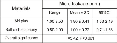 | Table 1: Evaluation of Apical Micro leakage.
 |
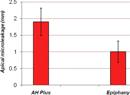 | Graph 1: Bar graph showing the mean level of apical leakage values of two groups.
|
STATISTICAL ANALYSIS
A one-way analysis of variance and post hoc tuckey test were used to seek statistically significant differences in apical leakage among the sealer groups.
RESULTS
The analysis of variance showed a statistically significant difference among the apical leakage of the two sealers (p < 0.001) demonstrating the influence of the type of sealer on the tightness of the apical filling. The result showed that the teeth filled with AH plus displayed a higher apical leakage (1.90 ± 0.41) than those filled with Self etch epiphany (1.00 ± 0.32).
Discussion
Preventing contamination or recontamination of the root canal system after completion of endodontic therapy is still a challenge for the dental professional. In the absence of impermeable sealing of the root canal system, failure of endodontic therapy may ensue.3
A superior method of sealing the apical foramen at the cemento-dentinal junction creates a favourable biological environment for periapical healing to take place by sealing of any communication between periodontium and root canal system.4 The complete seal of the root canal system is almost impossible with currently accepted materials and obturation techniques, usually a combination of Gutta Percha and zinc oxide eugenol root canal sealer is commonly adapted practice. Ideally, the root canal sealer should be capable of producing a bond between the core material and the root dentine, effectively preventing leakage. The ZnOE sealer lack properties required as root canal sealer. The present study was carried out to evaluate the apical sealing ability of newer resin based root canal sealers.5
Recent improvement in adhesive technology has led to the development of a new sealer, Epiphany that has a potential to challenge other sealers. Epiphany root canal sealant is a dual curable resin composite containing a new redox catalyst that enables optimal autopolymerization under acidic environments. The advantages of epiphany system includes high radiopacity, tissue compatibility, minimal shrinkage and resorbability of sealer when expressed periapically.6
In the present study, saline was used for the storage of freshly extracted teeth because it does not influence chemical and physical properties of human dentin.7
Single rooted premolars with single patent canals, were selected to minimize anatomical variation and allow standardization.5 Also the teeth were resected at cemento-enamel junction using water-cooled diamond disk to simplify instrumentation and obturation.8
The removal of smear layer may be considered an essential step in the process of successful root canal treatment. It is well known that root filling materials penetrate better into dentinal tubules in absence of smear layer. For this reason Smear layer was removed using 5.25% NaOCl and 17% EDTA to remove the smear layer and to evaluate the penetration and adaptation of root canal filling materials in the present study.5 However, sodium hypochlorite has been shown to adversely affect the bond strength of epiphany sealer to root dentin. Thus, EDTA is recommended to be used as final irrigant.9
All samples were coated with two coats of nail paint leaving apical 2 mm to prevent dye penetration through the root surface.10
The quality of apical seal obtained by root canal obturation material has been assessed by various methods like dye penetration, radioisotope penetration, bacterial leakage, fluorometric and electrochemical means, fluid filtration, scanning electron microscope and gas chromatography.11 The dye penetration was used because of its simplicity, ease to perform and it does not require sophisticated materials. Methylene blue dye was used as its molecular size is similar to bacterial by-products such as butyric acid which can leak out of infected root canals to irritate periapical tissues. 12
The root canals were evaluated using a sectioning technique. There are three advantages of sectioning technique compared to clearing technique namely, conservation of tooth substance for further analysis, considerably less time involved and lower costs.5
Results of the present study revealed that, Epiphany sealer exhibited the least apical leakage compared to AH plus. This may be attributed to the monoblock provided by adhesion of the filling material to the sealer which also adheres and penetrates into the dentin wall of the root canal system.12 The low mean apical leakage could be because of the attachment of the sealer to the root canal walls by its bonding agent and adhesion of the sealer to both the obturation material and to the dentin forming a monoblock.5
Under the conditions of our study, none of the materials produced an effective apical seal, and most leakage occurred between the wall of the root canal and the sealer. It is often stated that leakage may be influenced by the ability of a root-canal sealer to bond to the dentinal wall or at least to maintain permanent contact with the wall.10 In this respect we found that AH plus demonstrated maximum leakage and Epiphany exhibited the least which is similar to the studies by Emre Bodrumlu12 and F. Kont Cobankara2.
Though Epiphany system exhibits monoblock effect still leakage was observed, which may be attributed to inadverdent stripping of the sealer off the canal wall during placement of cones, disruption of the maturing resin-root dentin bond during cold lateral condensation or the C-factor.13 The manufacturer's instruction for immediate light-curing the coronal root filling to create a coronal seal may also limit flow of the resin sealer for stress relief.6
A study done by Tunga et al compared the sealing ability of Epiphany to AH Plus sealer and found significantly lower leakage with the Epiphany group due to increased adhesion of the sealer to the root canal walls which is in accordance with the present study.5
However, these findings are in contrast with the results obtained by Tay et al, where it was concluded that the quality of apical seal achieved by Resilon/Epiphany is not superior to Gutta Percha and conventional epoxy resin sealer. Discrepancies between the studies could be because of differences in methodology. 5
The mean leakage with Resilon/Epiphany system was lower than that for gutta-percha with AH plus sealer. The difference may be because of lack of bonding between gutta-percha and sealer.13 The sealing ability of AH Plus may also be affected by other factors; for example, AH Plus contains silicone oils and other ingredients. As all specimens were kept in 100% humidity one can speculate that oil-based materials such as AH Plus could prevent complete wetting of the root-canal wall and adhere poorly to humid dentine. This may result in poor adaptation of the material to the root-canal wall, as well as formation of voids that enhance dye penetration.14
The result in the present study showed that Epiphany provides a better consistent seal as compared to AH plus. However further long term studies both in vitro and in vivo with more variables are required for evaluating sealing ability of Epiphany.
Conclusion
Within limitations of our study AH plus is showing the highest microleakage while Epiphany is showing the least. Results of the present study should be interpreted with caution, and need to be investigated further.
References
1. Pommel L, Imad A, Pashley D, and Camps J. Apical Leakage of Four Endodontic Sealers. J Endod 2003; 29(3):208-210.
2. F. Kont C¸obankara, N. Adanir, S. Belli & D. H. Pashley. A quantitative evaluation of apical leakage of four root-canal sealers. Int Endod J 2002; 35: 979-984.
3. Kopper P, Vanni J, Bona A, Figueiredo J, Porto S. In Vivo Evaluation Of The Sealing Ability Of Two Endodontic Sealers In Root Canals Exposed To The Oral Environment For 45 And 90 Days. J Appl Oral Sci. 2006;14(1):43-48.
4. Rajput J, Jain R Lb, Pathak A. An Evaluation of Sealing Ability of Endodontic Materials as Root Canal Sealers. J Indian Sot Pedo Prev Dent March 2004; 22 (1): 1-7.
5. Aptekar A, Ginnan K .Comparative evaluation of microleakage and seal of two obturation materials Resilon/ Epiphany and Gutta Percha .J Can Dent Assoc 2006;72(3):245-249.
6. Franklin R. Tay et al. Ultrastructural evaluation of the apical seal in roots filled with polycaprolactone-based root canal filling material. J Endod 2005; 31(7): 514-519.
7. Gernhardt CR, Dr med dent, Kruger T, Bekes K, Schaller HG. Apical sealing ability of 2 epoxy resin-based sealers used with root canal obturation techniques based on warm gutta-percha compared to cold lateral condensation. Quinte Int 2007; 38(3): 229-234.
8. Lares C, Eldeeb ME. The sealing ability of the Thermafil obturation technique. J Endod 1990; 16(10):474-479.
9. Nunes HV, Silva RG, Alfredo E, Sousa-Neto MD, Silva-Sousa YTC. Adhesion of epiphany and AH plus sealers to human root dentin treated with different solutions. Braz Dent J. 2008; 19(1): 46-50.
10. Hata G, Kawazoe S, Toda T, Weine FS. Sealing ability of thermafil with and without sealer. J Endod 1992; 18(7):322-326.
11. D.M. Dalat, L.S.W. Spangberg. Comparision of apical leakage in root canals obturated with various gutta percha techniques using a dye vacuum tracing method. J Endod. 1994; 20(7): 315-319.
12. Bodrumlu E, Tunga U. Apical Leakage of resilontm obturation material. J Contemp Dent Practice 2006; 7(4):1-4.
13. Leonard JE, Gutmann JL, Guo IY. Apical and coronal seal of roots obturated with a dentine bonding agent and resin. Int Endod J 1996; 29:76-83.
14. Gençoglu N, Samani S, Günday M. Dentinal wall adaptation of thermoplasticized gutta-percha in the absence or presence of smear layer: a scanning electron microscopic study. . J Endod. 1993; 19(11):558- 562.
15. Zmener O. et al, Sealing properties of a new epoxy resin based root canal sealer, Int Endod J 1997; 30, 332-334. |