INTRODUCTION
Traumatic injuries most commonly affect maxillary incisors (80% central incisors and 16% lateral incisors) due to their anterior position and protrusion caused by the eruptive process.2 Gender also plays a major role on the incidence of traumas. It has been reported males are more frequently affected than females, particularly in the maxillary incisors.3,4
The treatment of complicated crown-root fractures in many cases is compromised by tooth fractures that are well below the gingival margin or bone. After root canal obturation, proper isolation for a dry operation field is critical for successful restoration of traumatized teeth.
Re-attachment of a tooth fragment should be preferable to restoring fractured teeth. There are several advantages in this treatment such as obtaining esthetic in a single appointment, being more conservative procedure, obtaining healthy periodontal attachment and it maintains the original tooth contours and translucence as the patient's own.5,6 The present case report describes the reattachment of an original tooth fragment using a fiber post.
CASE REPORT
A 10yr old male reported to the department of Pedodontics, Subharti Dental College with the chief complaint of fractured maxillary central incisors. Patient gave the history of trauma while playing cricket 1 day prior. The patient's medical history was non-contributory.
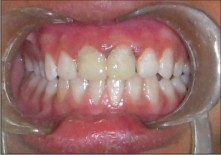
Examination revealed that the patient has fractured both maxillary central incisors [Figure 1].
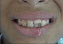 | Figure 1: Fractured Maxillary Central Incisors
 |
Oblique crown-root fracture in the maxillary right central incisor involved the enamel-dentin junction and extended from buccal to the palatal aspect subgingivally, and fractures in left central incisor involving the pulp.
After anesthesia, mobile coronal tooth fragment was removed. Full thickness buccolingual mucoperiostal flap was raised with an intrasulcular incision (Figure 2).
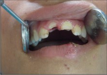 | Figure 2: Mucoperiosteal Flap Raised & Mobile Tooth Fragment Extracted # 11
|
Root canal pulp extirpation and canal preparation was performed using the standard step-back method. The prepared teeth were dried with paper-points and filled with laterally condensed gutta-percha and root canal sealer. After sealer set, the gutta-percha was partially removed from the root canal using heated instrument, leaving 5 mm of the filling material at the apex to maintain a good sealing. A post hole within root and coronal fragment was prepared using a drill recommended by manufacturer. A fiber post was cemented within root canal with dual adhesive cement (figure 3).
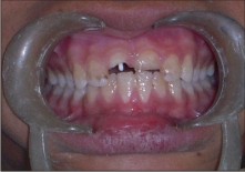 | Figure 3: Fiber Post # 11
|
After all debris on the fractured tooth surface was scaled, washed away with sterile serum physiologic and dried (Figure 4).
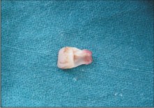 | Figure 4: Fractured Tooth
|
A primer was applied onto the fractured surfaces of the tooth fragments for 20 second. Volatile ingredients were evaporated with mild air stream. Bonding agent was applied to the primed surfaces of the fragments and light cured for 10 second. The fragments were reattached with a composite resin. The excess resin was removed with an excavator and the crown was light cured for 40 seconds from both buccal and palatal aspects. Final polishing of the crown root interface was made with ultrafine diamond burs and polishing disks. After all, the flaps were sutured. One week later, the sutures were removed and clinical examination revealed proper healing (Figure 5).
 | Figure 5: Post Operative (After 1 Week)
|
Patient was kept on regular follow up.
DISCUSSION
The fracture of a tooth may be a most traumatic incident for a young patient, but it has been found that there is a positive emotional and social response from the patient to the preservation of natural tooth structure6
Re-attachment of fragments offers a good esthetic, less time-consuming and cost-effective restorative option.7 It is conservative, has favorable wear mechanism, helps to preserve occlusal contacts and is well accepted by the patient.8
Some of the disadvantages of reattachment includes: the necessity for continuous monitoring, its unknown longevity, and possibility of color changes of the bonded fragment.8
Andreasen and Andreasen state that the reattachment procedure may importantly serve as a transitional treatment alternative for pre-teens or teenage patients to postpone definitive treatment until an age where gingival margin contours are relatively stable .9
Liew believed this restoration to act as " a short to medium term temporary restoration which has the potential for indefinite service" .9
A post and core is needed to improve retention, to distribute stress and to improve resistance to root fracture. The post interlocks the two fragments and minimizes the stresses on the remaining tooth structure that is replaced.8
Fiber-reinforced composite resin post has demonstrated negligible root fracture. In addition, the fiber-reinforced posts can be used with minimal preparation because it uses the undercuts and surface irregularities to increase the surface area for bonding. Thus, it reduces the possibility of tooth fracture during function or traumatic injury.10
Reattachment of the dental fragment as a restorative procedure becomes possible with the improvement of adhesive techniques and restorative materials.6
A multicenter clinical study done by Andreasen FM et al concluded that the good fragment retention , acceptable esthetics, and pulp vitality indicated that reattachment of coronal fragment is a realistic alternative to placement of conventional resin composite restoration.11
The reattachment technique was first described in 1964 by Chosak and Eidelaman, at that time, it was considered as a provisional restoration due to the low bond strength values achieved by the adhesive systems. However, the remarkable advancement of the adhesive systems and resin composites has made the reattachment of tooth fragments a procedure that is no longer a provisional restoration, but rather a treatment offering favorable prognosis. This procedure found a strong argument in a conservative philosophy, since it does not require excessive wear of the healthy tooth structure and do not make unfeasible any other later possible restorative treatment.12
The re-attachment of a tooth fragment is a viable technique that restores function and esthetics with a very conservative approach, but for each trauma case should be attempted to restore on an individual basis.
CONCLUSION
Reattachment of the intact fractured segment can be considered as an ultraconservative method for aesthetic rehabilitation. This procedure helps us to preserve maximal natural tooth structure. The superior quality adhesive materials make this procedure viable. The need of the day is to educate the population to preserve the fractured segment and seek immediate dental treatment.11
REFERENCES
1. Ozel E, Cildir A, Ozel Y. Re-attachment of Anterior Tooth Fragment using a Self-etching Adhesive:A Case Report. J Contemp Dent Pract 2008 January; (9)1:77-083.
2. Andreasen JO, Andreasen FM. Textbook and color atlas of traumatic injuries to the teeth Munksgaard, Copenhagen, 1993.
3. Forsberg CM, Tedestam G. Etiological and predisposing factors related to traumatic injurities to permanent teeth. Swed Dent J 1993;17:183-190.
4. Rapelli G, Massaccesi C, Putignano A. Clinical procedures for the immediate reattachment of a tooth fragment. Dent Traumatol 2002;18:281-284.
5. Arhun N, Ungor M. Re-attachment of a fractured tooth: a case report. Dent Traumatol 2007; 23:322-326.
6. Baratieri L.N., Monteiro S.: Tooth fracture reattachment: Case reports. Quint Int 1990; 21: 261 - 270.
7. Zorba YO, Ozcan E. Reattachment of coronal fragment using fiber-reinforced post: a case report. Eur J Dent 2007; 1:174-178.
8. Kavitha T, Rao CVN, Lakshmi Narayan L: Reattachment of fractured tooth fragments using a custom fabricated dowel- Three case reports Endodontology , vol 12,2000.
9. D.F. Murchison et al; Incisal edge reattachment: indications for use and clinical technique; british dental journal, vol 186(12),june 1999: 614-619.
10. Yahya orcun Zorba, Erdal Ozcan: Reattachment of coronal fragment using fiber-Reinforced post: A Case Report; Eur J Dent. 2007 July; 1(3):174-178.
11. Thejokrishna.P,Prabhakar AR,Kurthukoti AJ : Reattachment Of Embedded Tooth Fragment: A Case Report. Annals and essence of dentistry, 2010
12. Roberta Caroline Bruschi ALONSO et al: Reattachment of an autogenous tooth fragment - 36-month follow-up: fast and safe rehabilitation of fractured teeth; Perspect. Oral Sci. | v.1 | n2 | DEZ | 2009.
13. Joshi N, Shetty N, Kundabala M: Immediate reattachment of fractured tooth segment using dual cure resin; Kathmandu University Medical Journal (2008), Vol. 6, No. 3, Issue 23, 386-388. |