|
|
|
| An Unusual Eruption Of Mandibular Third Molar At The Age Of 64 Years : A Case Report |
Rakesh Kumar Manne 1
1 M.D.S. (Asst. Professor,) - Department of Oral Medicine & Radiology, Vyas Dental College & Hospital
|
| Address For Correspondence |
Dr. Rakesh Kumar Manne. M.D.S
Asst. Professor,
Department of Oral Medicine & Radiology
Vyas Dental College & Hospital,
Pali road,Jodhpur, Rajasthan, India.
Phone: 09680974277, 09704014747
E-mail: rmannae@rediffmail.com |
| Abstract |
| The eruption of the third molars occurs between the ages of 18 to 20 years. The eruption at a time that deviates significantly from normal eruptive ages is delayed tooth eruption. In this report, an unusual case of delayed right mandibular third molar eruption at the age of 63 years is described. Also discusses the diagnosis and possible reasons of this unusual tooth eruption. |
|
| Keywords |
| Age, Eruption, Mandible, Molars |
|
| Full Text |
INTRODUCTION
Tooth development begins in the fetus at about 28 days in uterus, indeed, all the primary and some of the permanent dentition start to develop in the fetus. Mineralization of the primary dentition commences at about 14 weeks in uterus, and all primary teeth are mineralizing by birth. Tooth eruption occurs after formation and mineralization of the crown are largely complete but before the roots are fully formed (1). The Complete eruption of the third molars occurs between the ages of 18 to 20 years. The mandibular third molars erupt generally ahead of maxillary third molars (2).
A delay in third molar eruption of up to 12 months beyond normal eruptive age may be of little or no importance in healthy individuals. The eruption at a time that deviates significantly from normal eruptive ages is delayed tooth eruption. Delayed third molar eruption may or may not have identifiable local or systemic causes. The identifiable local factors includes tooth in the path of eruption, insufficient space in the dental arch, dental infection, ectopic positions of teeth, supernumerary teeth etc. Systemic and genetic factors like cleidocranial dysplasia, osteopetrosis etc. may also cause delayed eruption (3).
CASE REPORT
A 65 yrs old male farmer reported to the department of Oral Medicine and Radiology with a complaint of missing all the teeth for the past 2 years and wants a complete denture (Fig. 1).
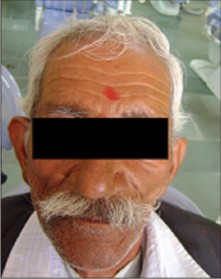 | Fig. 1: Extraoral photograph showing geriatric status of the patient
 |
The history of present illness revealed all the teeth where extracted due to mobility and lower right back tooth was the last tooth to extract. A new tooth was erupted 4 months after the last extraction at the same site. Patient wants a denture with out extracting the newly erupted tooth. There was no associated medical history and patient had a normal physical and mental health.
Intraoral examination showed all the teeth where missing except right mandibular third molar. Right mandibular third molar crown appears bulbous with prominent cuspal pattern, shows evidence of occlusal pit and fissure caries and slightly extruded from the socket (Fig. 2).
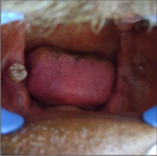 | Fig. 2: Intraoral photograph showing right mandibular third molar with bulbous crown, prominent cuspal pattern, and occlusal pit and fissure caries
|
No clinical evidence of age related changes in tooth or periodontium is seen. Intraoral periapical radiograph shows normal cuspal pattern, normal pulpal dimensions and single root in right mandibular third molar. Radiograph also shows no periodontal bone loss and healing extracted tooth socket of right mandibular second molar (Fig. 3).
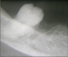 | Fig. 3: Intraoral periapical radiograph showing normal cuspal pattern, normal pulpal dimensions, and single root in right mandibular third molar
|
Considering the patient's age, history of present illness, clinical, and radiographic findings a diagnosis of delayed eruption of right mandibular third molar was made.
DISCUSSION
Dentition and its supporting periodontium undergo age related changes. Dental age related changes are tooth discoloration, loss of enamel due to attrition, abrasion and erosion (4), increased caries susceptibility, cemental thickness (5), and reduced pulp dimensions (6). Age related immunological changes in the periodontal tissue could alter the host response to microorganisms, affecting the patient's ability to respond to periodontal treatment. Irregular dental visits and poor socioeconomic status are predictors for periodontal disease (7). The present patient is 65 years old from poor socioeconomic background showed no age related changes apart from the initial pit and fissure dental caries suggestive of recently erupted tooth.
Several theories have been postulated to explain the mechanism by which a tooth moves through its surrounding tissues as it erupts into the oral cavity. The most acceptable are root formation, bone remodeling, and periodontal ligament formation and renewal theories. Other suggested causes are periodontal ligament hydrostatic pressure, the activity of the pulp, including constriction by dentinogenesis and increase in the volume area of Hertwig's epithelial root sheath and the diaphragm, pressure exerted by muscles, and hormonal influence, particularly by pituitary and thyroid hormones (8). Root formation theory states overall increase in the length of the tooth may be accommodated by the growth of the root into the bone of the jaw, by an increase in the jaw height, or by the occlusal movement of the crown. The evidence of rootless tooth eruption, tooth erupting greater distance than the total length of their roots, and the tooth erupting after the completion of root formation discarded the root formation theory of tooth eruption (9). In the present case we also discard the root formation theory in the eruption of right mandibular third molar because root formation of right mandibular molar would have completed long back before eruption and tooth still erupted.
Bone remodeling theory states the inherent growth pattern of the mandible or maxilla supposedly moves tooth by the selective deposition and resorption of bone in the immediate neighborhood of the tooth. The bone remodeling is a continuous process. The rate of bone remodeling comes down but does not stop with age (9, 10). Bone remodeling could be one of the reasons for the eruption right mandibular third molar in the present case because bone remodeling is a continuous process even at the age of 65. Periodontal ligament formation and renewal theory states the constant turnover of collagen fibers in the periodontal ligament. During maturation, collagen fibers shrink in length by about 10%.because of the orientation of these oblique collagen fibers, the vector force generated in occlusal direction results in tooth eruption. It also states a small, but measurable, contractile force can be generated by fibroblasts. Because fibroblasts are the most numerous cell types in the periodontal ligament, and they can attach to collagen type 1 fiber via fibronectin and integrins (11). Periodontal ligament formation and renewal can occur through out the life and could also be the reason for the eruption of mandibular third molar in the present case.
CONCLUSION
No case report of delayed tooth eruption at the age of 64 has been reported in the literature till today. No one theory of how the forces might be generated to cause tooth movement has been put forward that can account for all aspects of eruption. Tooth eruption is probably multifactorial in that more than one mechanism may be involved. In the present case we assume mandibular bone remodeling and periodontal ligament formation and renewal mechanisms would have played a possible role in the eruption of right mandibular third molar. Further studies needs to be carried out to understand the exact mechanism of tooth eruption.
REFERENCES
1. Ruth Holt, Graham Roberts, Crispian Scully. Oral health and disease. West J med 2001; 174: 199-202.
2. S A Odusanya, I O Abayomi. Third molar eruption among rural Nigerians. Oral Surg Oral Med Oral Pathol 1991;71:151-154.
3. Fatih Ozan, Isa Kara, Sinan Ay. Impacted Mandibular Permanent Incisors Associated With a Supernumerary Tooth: A Case Report. Eur J Dent 2009;3:324-328.
4. Whittaker DK, Bakri MM. Racial variations in the extent of tooth root translucency in ageing individuals. Arch Oral Biol 1996;41:15-19.
5. Ketterl W. Age-induced changes in the teeth and their attachment apparatus. Int Dent J 1983;33:262-71.
6. Morse DR, Esposito JV, Schoor RS. A radiographic study of aging changes of the dental pulp and dentin in normal teeth. Quintessence Int 1993;24:329-33.
7. Axelsson P, Paulander J, LindheJ. Relationship between smoking and dental status in 35-, 50-, 65-, and 75-years old individuals. J Clin Periodontal 1998;25:290-8.
8. Rakhi Gupta, B Sivapathasundharam, A Einstein. Eruption age of permanent mandibular first molars and central incisors in the south Indian population. Indian J Dent Res 2007;18:186-189.
9. Nanci A (ed). Tencate's oral histology. Elsevier: India,2008:271-73.
10. Parfitt AM. The bone remodeling compartment: a circulatory function for bone lining cells. J Bone Miner Res 2001;16:1583.
11. Gaunt WA, Osborn JW, Tencate AR (ed). Advanced oral histology. John Wright and Sons Limited: Bristol,1971:114-7. |
|
|
|
|
|
|