Introduction
Main purpose of post and core restoration is to provide the substructure to which the final restoration can attach and anchor the root.1 Posts are required to support a core foundation when there is insufficient remaining structure of clinical crown.2 following post and core systems are commonly employed to restore the endodontically treated teeth.
Traditionally, the cast post and core has been the means of restoring these teeth.3 Cast posts are made of stainless steel, Nickel-chromium, Pure Titanium, Titanium alloy or non oxidizing noble alloy. One disadvantage of cast post and core is that cast post concentrates the stresses closer to the post itself. This may result in post failure and root fracture. Prefabricated post systems are popular because they save time and can provide satisfactory results. A variety of prefabricated post systems have been introduced over the years and used successfully in clinical situations.3The growing demand for esthetic restorations in dentistry has led to the development of tooth-colored, metal-free, dowel and- core systems.4 Fiber post composite restorations improve tooth flexibility under applied load as well as stress distribution between post and dentin to provide esthetic means of building up a post and core at chair side.5
Comparative analysis of fracture strength of different post and system employed to restore restore maxillary central incisors with severe horizontal defect seems to be lacking. Thus this In vitro study was undertaken using a universal testing machine to evaluate and compare the fracture strength of glass fiber reinforced composite post and core, Titanium alloy post and composite core, and custom cast Ni-Cr post and core system, used to restore maxillary central incisors with severe defects.
Material and Method
This study was conducted in the Department of Prosthodontics Crown and Bridge HDC Sundernagar on thirty, freshly extracted human maxillary central incisors. The teeth were collected irrespective of the age, sex, and side of the arch. All the teeth were without root caries, root fillings, root cracks, and minimum of 10mm of root length. The selected teeth were stored in artificial saliva (wet mouth ICPA C09002) at room temperature until used for study, to avoid their dehydration. All the teeth were decoronated horizontally at cemento-enamel junction, perpendicular to long axis of teeth. Endodontic treatment of selected teeth was completed as: A#15-50 using step back technique. Thirty teeth were prepared and all the prepared teeth were randomly divided into three groups of ten teeth each, as shown in Table I.
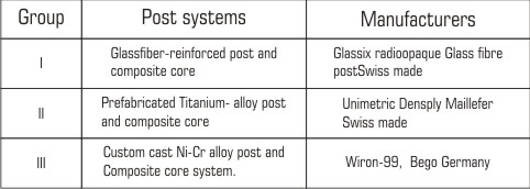 | Table I : Post Systems and their Manufacturers
 |
For group I (GFRP) and group II (Ti P), special drills supplied with the kit, were used to prepare post space, leaving 3 mm apical seal in the root canal. For group III (CP) Peeso reamers (#1-6, Mani Japan) were used to prepare the post space. Standardization was achieved with the help of rubber stoppers placed on special drill and peeso reamers. After preparation of each post space a radiograph was taken to evaluate the length of prepared post space and remaining apical seal in the canal.
The posts were cemented with adhesive resin cement (3M) ESPE (N120174). Then it was light cured for 40 sec to achieve complete polymerization.
Core build up was done with Tetric N-cerem, Ivoclar Vivadent (N-12096). The preformed polyester matrix was filled with the core build up material in increments of 2 mm and placed on the specimen. Each increment was light cured for 60 sec. It was finished to the final core height of 6 mm and bucco-lingual and mesio-distal dimensions corresponding to that of the tooth, with the help of composite finishing kit.
For group III - AL paper pin was used to prepare a wax pattern of prepared post space. Paper pin was roughened and dipped in molten blue inlay wax and inserted into the canal. Incremental addition of wax was done to make a post pattern. The core was built up to achieve a desired core height of 6 mm, and bucco-lingual and mesio-distal dimensions corresponding to the total bucco-lingual and mesio-distal dimensions of the specimen. The post and core wax pattern was sprued and invested, and casting of post and core were obtained. Etching bonding and cementation of cast post were done as same procedure was done for group I and group II post system. With free hand all preparations were finished with a diamond bur (DIA BURS,WR-13 ISO 068/042) at high speed with water spray(W&H Dentalwerk Burmose Gmbh Austria). All finish lines were placed at level of cemento-enamel junction. Single coat of spacer was applied to the core part of the specimen. The core was dipped into the molten blue inlay wax, to give a uniform layer of wax over the entire surface. Crown patterns were obtained on all three groups of post and core groups by duplicating polycarbonate crown forms (size 10).Investing and casting was done as same procedure was done above. Castings were retrieved, finished and polished
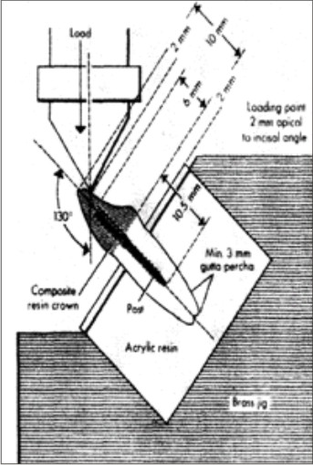
Cementation of crown was done with the adhesive resin cement. All specimens roots and were dipped into molten wax to a depth of 2mm below the CEJ to give a uniform layer 0.2-0.3 mm of wax over the entire root surface.30 Acrylic resins blocks were fabricated by mixing the self- cure acrylic resin DPI-RR cold cure P-13103, L-13102 and pouring it into block - holder part of metal jig. Specimens were held perpendicular into the center of block - holder part of metal jig. After setting of acrylic resin, dewaxing was done to achieve a space for simulated periodontal ligament. Type I- regular viscosity regular body, polyvinyl siloxane impression material (Reprosil-Dentsply) 100313 was applied over the root surfaces and also into the mold surface in acrylic resin blocks. Each specimen was reinserted into acrylic blocks upto2mm below CEJ and held under digital pressure until material was set. Excess material was removed with the help of B. P. knife. All specimens were prepared as seen in fig (1)
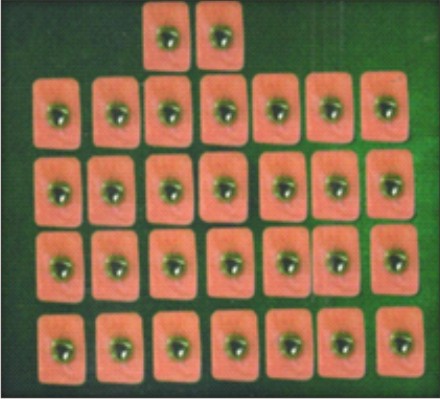 | Fig 1. Specimen Prepared For Testing
|
and again stored in artificial saliva until tested. Stainless steel metal attachment tool were custom made of the desired dimensions according to the testing machine. Metal jig had the provision for holding acrylic resin blocks that orient the specimen at an angle of 130 degree to the load application tip of the attachment tool. The whole assembly was fitted in the universal testing machine and load was applied on the palatal surface of cast crown, 2mm from incisal edge, at an angle of 130 degrees to the long axis of the root, at a cross head speed of 1mm\min until failure occurred as seen in fig 2(a,b).
 | Fig 2(a). Testing Assembly
|
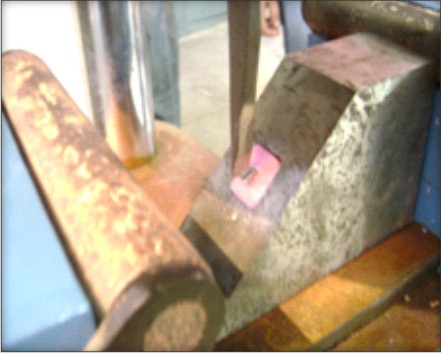 | Fig 2(b). Testing Specimen On Universal Testing Machine
|
Observations and Results
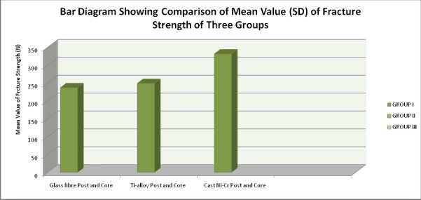
The mean value for fracture resistance was (233.24±26.20) N, (249.00±31.55)N, (330.39±30.81)N in group I Glass fiber reinforced composite post and core, group II Titanium alloy post and composite core, group III custom cast Ni-Cr post and core) respectively shown in Chart I.
The custom cast Ni-Cr post and core specimens presented the higher mean values of fracture resistance as compared to group I and group II. Analysis of Variance (one way ANOVA) of fracture resistance results shows the significant difference amongst the three groups P<0.01 (Table II).
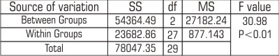 | Table II : Analysis of Variance (one way ANOVA) of fracture Strength
|
It was observed that the intergroup comparison was highly significant for group I (GFRP) & (CP), (t-value 7.59 at p-value <0.001); and Group II (Ti P) & (CP), (t-value 5.83 at p-value <0.001).The difference was non significant for group I (GFRP) & II (Ti P) as shown in table III.
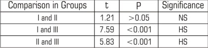 | Table III : Comparison of Variance of fracture Strength
|
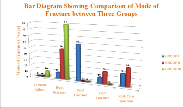
In Group I - Glass fiber reinforced composite post and core system (GFRP), no specimens showed cement failure, 60% of the specimens had post fracture, root and core fractures occurred in 10% of the specimens. In Group II - Titanium alloy post and composite core system (TiP) no specimens showed cement failure and post fracture,
50% of the specimens had the root fracture, 30%of the specimens had post-core junction fracture and 20% of the specimens had core fractures. In Group III - Custom cast Ni-Cr post and core system (CP), 10% of the specimens had cement failure, 90% of the specimens had root fracture. Post fracture, core fracture and post-core junction fractures were not seen in any specimen. (As shown in Chart II)
Discussion
Thirty freshly extracted human maxillary central incisors were taken for the study. All teeth were decoronated at the cemento-enamel junction to simulate the commonly encountered clinical situations of lost tooth structure.The radicular portion of each tooth was embedded in rapid repair acrylic resin 2 mm below the proximal cemento -enamel junction. This simulated the clinical biological distance. The position of the central block directed the load at an angle of 130 degree to central long axis of root. This configuration provided an even distribution of forces along the perpendicular axis of the root and also replicated flexion stress resulting from protrusive movements. This angle was selected due to its proximity to the ideal 134.5 degree inter-incisal angle observed in angles class I occlusal relationship.6, 7
Teeth restored with cast post and core system exhibited significantly higher fracture resistance followed by titanium and glass fiber post and core system. This can be attributed to the fact that cast post has better physicomechanical properties in comparison to titanium post and glass fiber post system. 8
This is in agreement with studies conducted by Insua et al, Sidoli GE et al, Sirimai S et al and Eskitascioglu et al 8, 9, 10, 11. In the second part of the study the mode of failure of various post systems were observed. The various mode of failure observed were (a) cement failure (b) Root fracture (c) Post fracture (d) Core fracture
(e) Post-core junction fracture
The incidence of cement failure is not significant among the three groups. The cement used in this study was resin adhesive cement. This cement provides stronger union between post and core and tooth structure using adhesive bonding technique. It has been claimed by Mohhamad N12 this adhesive restorative cement transmits and distributes functional stresses across the bonding interface to the tooth more properly with the potential to reinforce weekend tooth structure. According to Freedman GA13, resin based composite is an esthetically pleasing materials which has good strength. Cormier CJ14 described that integration of adhesive technique into post and core procedures resulted in "monobloc" type of restoration.
The prevalence of root fracture was highest in case of cast post system followed by titanium post and glass fiber post system. This is in agreement with studies conducted by Insua et al 8, Sidoli GE et al9, Sirimai S et al.10 This can be explained by the fact that cast restorations have highest modulus of elasticity (220 GPa) as compared to titanium post (112 GPa) and glass fibre post (13-40 GPa).whereas the modulus of elasticity of dentine is (15-25 GPa). The high difference in modulus of elasticity as compared to dentin has resulted in catastrophic root fractures in case of cast post and titanium post systems. Due to the near comparable modulus of elasticity to dentin in case of glass fiber post the incidence of root fracture is less.
The findings of Chart II regarding the incidence of post and core fracture are insignificant. A few Incidence of post fracture are observed in glass fiber post and core system. This can be due to the weak bond between the internal glass fibres and resin matrix 15. The composite core fracture is more prevalent in case of titanium post system as compared to glass fibre post and composite core system. The solid resin core specimens had the highest mean DTS values whereas cores with metallic post groups had the lowest mean DTS value.15
High incidence of post and core junction fracture is observed in case of titanium post and core system followed by glass fibre post and cast post system as shown in Chart II. This can be explained on the basis of the fact that in cast post system cast metal resisted the force and transferred it to the root16. For metallic post specimen failure occurred at the interface between the post surface and the resin composite material, with the cohesive strength of both materials being higher than that of the interface. However with nonmetallic post the failure predominantly occurred along the interface between the resin coating at the surface of the post and the reinforcing fibers comprising the majority of post15.
Drawback of this study was that it was an in vitro study and results obtained may not be comparable to in vivo situations. Some factors such as quality and quantity of remaining tooth structure can explain the variations in the result. Therefore further studies may be conducted including the above mentioned criteria.
Summary and conclusions
Within the limitations of the present study the following conclusions can be drawn:
1. Significantly higher fracture resistance was recorded in the group III custom cast post system than teeth restored with other two systems.
2. The titanium post system showed less fracture resistance as compared to teeth restored with custom cast post and core system and the most catastrophic failures.
3. A more favourable mode of failure was observed in teeth restored with Group I glass fibre post system.
4. Teeth restored with group III custom cast post system showed catastrophic vertical root fractures which are non repairable.
References
1. Cohen BI, Musket BL, Deutsch AS. Comparison of retentive properties of four post systems. J Prosthet Dent 1992; 68:264-268.
2. Valle AL, Pereira JR and Shiratori FK. Comparison of the fracture resistance of endodontically treated teeth restored with prefabricated posts and composite resin cores with different post lengths. J Applied Oral Sciences. 2007; 15(1):29-32.
3. Volwiler RA, Nicholls JI and Harrington GW. A comparison of three core build-up materials used in conjunction with two post systems in endodontically treated anterior teeth. Journal of Endodontics 1989; 15: 355-361.
4. Akkayan B. An in vitro study evaluating the effect of ferrule length on fracture resistance of endodontically treated teeth restored with fiber-reinforced and zirconia dowel systems. J Prosthet Dent 2004; 92:155-62.
5. Arcangelo CD, Angelis FA, Vadin M, Zazzeroni S, Ciampoli C and D'Amario M. In vitro fracture resistance and deflection of pulpless teeth restored with fiber posts and prepared for veneers. J Endod 2008; 34:838 - 841.
6. M. Sadeghi. A comparison of the fracture resistance of endodontically treated teeth using three different post systems. Journal of Dentistry, Tehran University of Medical Sciences, Tehran, Iran 2006; 3:69-76.
7. Federick DR. An application of the dowel and composite resin core technique J Prosthet Dent 1974; 32:420-5.
8. Insua MA, Silva LA, Rilo B, and Santana U. Comparison of the fracture resistances of pulpless teeth restored with a cast post and core or carbon-fiber post with a composite core. J Prosthet Dent 1998; 80:527-32.
9. Sidoli GE, King PA and Setchell DI. An in vitro evaluation of a carbon fiber-based post and core system. J Prosthet Dent 1997; 78:5-9.
10. Sirimai S, Riis DN, and Morgano SM. An in vitro study of the fracture resistance and the incidence of vertical root fracture of pulpless teeth restored with six post-and-core systems. J Prosthet Dent 1999; 81:262-9.
11. Eskitasc?oglu G, Belli S and Kalkan M. Evaluation of two post core systems using two different methods fracture strength test and a finite elemental stress analysis. Journal of Endodontic 2002; 28:629-633.
12. Mohammadi N, Kahnamoii MA, Yeganeh PK, and Navimipour EL. Effect of fiber post and cusp coverage on fracture resistance of endodontically treated maxillary premolars directly restored with composite resin. J Endod 2009; 35:1428-1432.
13. Freed man GA. Esthetic post and core treatment. Dent Clin North Am 2001; 45:103-15.
14. Cormier CJ, Burns DR and Moon P. In vitro comparison of the fracture resistance and failure mode of fiber, ceramic and conventional post systems at various stages of restorations .J Prosthodont 2001; 10:26-36.
15. Santos GC, El-Mowafy O and Rubo JH. Diametral tensile strength of a resin composite core with non-metallic prefabricated posts: an in vitro study. J Prosthet Dent 2004; 91:335-41.
16. Darabi F, Namazi L. Comparison of the fracture resistance of endodontically treated teeth using two different restoration systems. Dent Research Journal 2008; 5(2):65-69. |