Introduction
A precise knowledge about the physical and histological changes taking place in the dental hard and soft tissues due to extreme heat may prove to be of significance in ‘fire investigations’. In such investigations it is of paramount importance to find out the cause of fire, the exact location where the fire originated and the identification of the victims. Dental evidences may provide a clue to solve the mystery of victim identification, since dental structures are the last to get destroyed under extreme conditions, be it temperature, acid or putrefaction.
Materials and Method
Freshly extracted permanent teeth fixed in 10% formalin were collected and categorized according to the age of the patient. The group A comprised of teeth from patients less than 30 years, the group B comprised teeth from patients aged 30-40 years, and the group C comprised teeth from patients aged above 40 years.
Each group included 18 anterior, 18 posterior and 6 teeth restored with silver amalgam. Grossly decayed teeth which had very little hard tissue left were excluded.
A custom made steel tray that could withstand high temperatures (manufactured by Hebich Technical Training Institute, Mangalore.) was used (Fig.1a)
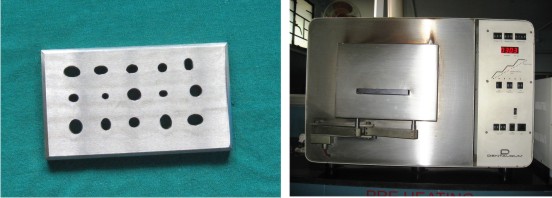 | Fig.1 : a. Custom-made Stainless steel tray, b. Electrical Furnace
 |
to keep 3 anterior, 3 posterior and one restored teeth in the upright position in the furnace. The varying sized holes in the tray held all teeth in place throughout the heating process.
The tray with teeth in position was placed in the electrical furnace (Fig.1b) and was subjected to a specified temperature for fifteen minutes, followed by gradual cooling to room temperature. The temperatures specified were 3000C, 3500C, 4000C, 4500C, 5000C and 6000C. The heating process involved gradual heating of teeth to the specified temperature, and holding the temperature for fifteen minutes, followed by gradual cooling to room temperature.
Teeth were then examined for any physical changes that occurred. These changes included change in color, texture or morphology. After a thorough physical examination teeth were placed in 10% formal formic acid for decalcification following fixation in 10% formalin. The decalcification fluid was changed every day, and the teeth were observed and documented for any significant physical changes during the process. Sponginess of dentin was considered to be the end-point of decalcification. Following complete and careful decalcification the tissues were kept for routine processing and were embedded in paraffin. Sections of 4 ? thickness were taken and stained in hematoxylin and eosin (H & E).
Result
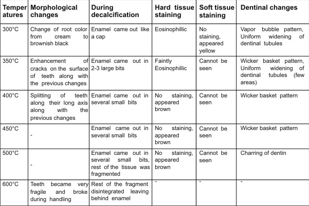
The morphological changes observed included change in color of root surface from white to brown, development of cracks, and splitting of teeth along its long axis. Teeth progressively became fragile and difficult to handle with increase in temperature (Fig 2a,b).
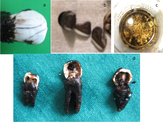 | Fig 2 : a. Change in color of root and enhancement of cracks, b. Splitting of teeth along long axis and black dicoloration of dentin, c. Disintegration of teeth in decalcification fluid after a thermal insultat 600 c, d. Detachment of Enamel cap along the
|
Teeth from all three groups which were subjected to a temperature of 6000C disintegrated completely as soon as they were put in the decalcification fluid. Therefore, processing and sectioning of these teeth could not be carried out. (Fig 2c)
Teeth subjected to lower temperatures demonstrated the following trends during the decalcification procedure. The teeth from group A, decalcified faster when compared to other two groups. The enamel caps of the teeth from all three groups separated out from the underlying supporting dentin along the dentino-enamel junction, before enamel could completely decalcify (Fig 2d). The enamel cap became increasingly fragile and came out in bits at temperatures greater than 300OC, separation still being at DEJ.
On examination of H & E stained sections it was noticed that the stainability of the tissues reduced progressively with increase in temperature. At 3000C soft tissues appeared yellow though dentin and cementum could be visualized clearly as taking up eosin stain (Fig 3a, b).
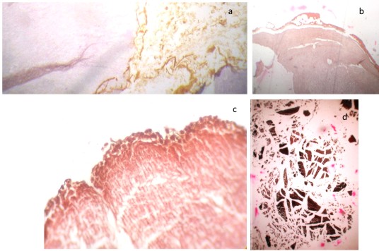 | Fig 3 : a. Pulpal tissue appearing yellow and dentine appearing faintly eosinophilic on H & E staining after treatment at 3000C, b. identification if dentine and cementum in teeth treated at 3000c,
|
One of the sections revealed remnants of dislodged amalgam filling on the surface of dentinal tubules in the form of spherical brown globules (Fig 3c). Dentin demonstrated irregular dilatations of tubules, that could be visualized in both longitudinal and cross-sections of dentin giving rise to a pattern referred 'vapor bubble pattern' (Fig 4a). Another interesting finding was the presence of uniform splitting of along the transverse axis of dentinal tubules. The thickness of each segment was found to be 15-20µm as measured by micrometer eyepiece reticule 1.
Histological changes perceived at 3500C included brown appearance of dentin and cementum with inability to differentiate between these two structures. Soft tissues could not be witnessed at and above this temperature. At this temperature dentine showed uniform widening of tubules that could be appreciated better in the cross-section of dentinal tubules (Fig 4b).
Dentin heated to 4000C and 4500C showed cohesion of dentinal tubules with splitting between the clusters giving rise to 'wicker basket pattern' (Fig 4c).
Teeth subjected to 5000C temperature were ineffectual for histological study and appeared charred (Fig 4d).
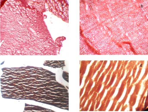 | Fig 4 : a. Vapor bubble pattern, b. Uniform widening of dentinal tubules, c. wicker basket pattern, d. charring of dentin
|
Discussion
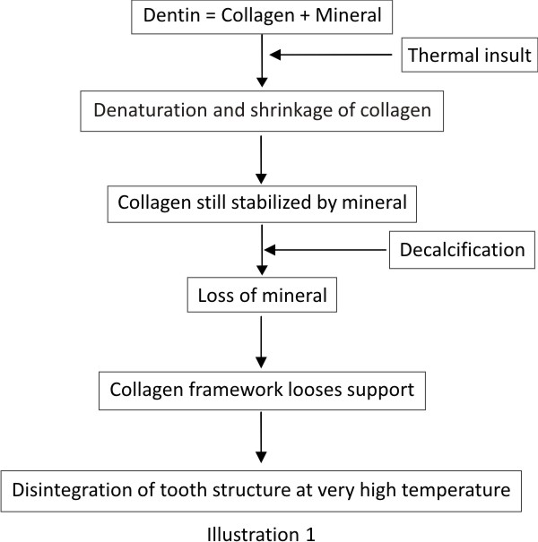
Enamel is made up of 96% mineral, where as dentin is composed of 76% minerals, and 20% organic component, mainly type 1 collagen 2. Mineral content helps stabilize collagen in dentin against thermal denaturation and shrinkage. But after decalcification minerals are lost and only a framework of collagen is left behind, which is destroyed at higher temperatures leads to disintegration of teeth in decalcification fluid 3. The higher the temperature more is the destruction of collagen framework and more is the disintegration of tissue in decalcification fluid (Illustration 1).
Possible explanation for the widening of dentinal tubules: Heat may lead to the formation of intratubular water vapor, which in turn may cause widening of dentinal tubules. If this is the case then similar changes should be seen even at any temperature between 1000C to 3000C.
Though temperature below 3000C is not encountered in cases of fire disasters or crimes, the study of teeth heated to this temperature may reveal some interesting observations.
Possible explanation for Wicker basket pattern: this pattern may appear due to a combination of three changes that heating may induce- Loss of inorganic matrix from decalcification, shrinkage due to drying and, incineration and consequent loss of intertubular organic matrix.
The presence of uniform splitting along the transverse axis of dentinal tubules at a distance of 15-20µm was consistent with the distance between incremental lines of Von Ebner seen in dentin. This finding emphasized the fact that incremental lines indeed are the weakest areas in hard tissues 4.
Teeth surviving decalcification were soft and fragile and difficult to section. Impregnation in gelatin can be used to provide better results 5.
Faster decalcification of younger teeth could be due to the fact that mineral content of younger teeth is lesser when compared to older teeth, as mineral content increases with age 2.
 |
|
 |
|
Conclusion
Routine histological evaluation of incinerated dental remains may provide additional investigative avenues both in the areas of fire investigation and victim identification. A larger sample size, more temperatures and variables like caries, restorations, and RCT treated teeth will provide a more reliable data for practical implications.
References
1. Richard Ten Cate ; Oral Histology: Development, Structure & Function ; 6th edition ; Mosby
2. Cellular Pathology Technique: C.F.Culling, R.T.Allison, W.T. Barr: 4th edition.
3. Orban's Oral histology & embryology ; 11th edition ; Mosby
4. The Microscope, Its Theory and Application: J.H.Wredden: 1st edition.
5. Myers SL, William JL, Hodges JS. Effects of extreme heat on teeth with implications for histologic processing. Journal of Forensic Sciences. 1999; 44 (4): 805-8 |