INTRODUCTION
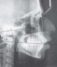
In 1931, Broadbent in the USA and Hofrath in Germany introduced the technique of radiographic cephalometry. Since then, clinicians and researchers have adopted and routinely used this valuable tool to analyze the underlying dentofacial relationships. Although the lateral cephalogram provides us a lot of information regarding the craniofacial structures, it is
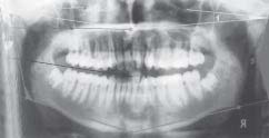
impossible to accurately visualize the right and left sides of these structures in a single radiograph due to the superimposition of the two sides. The panoramic radiograph developed by
Paatero. Y.V. in 1948 however allow the visualization of left and right sides of craniofacial structures by producing an accurate, predictable image of all the teeth and related structures on a radiograph, in the shortest possible time, with the least amount of radiation to the patient and to the operator and with the minimal amount of superimposition of various structures. Facial and
mandibular asymmetries are of special interest to the orthodontist. There are some published data about the effect of orthodontic treatment on the temperomandibular joint screened on the panoramic radiographs.
Aiming to enhance the clinical versatility of the panoramic radiograph, the objective of the present study is to investigate whether we can use panoramic radiograph instead of lateral cephalogram to assess dento skeletal pattern. In the present study, measurements will be taken from 60 panoramic radiographs and will be compared with the measurements taken from lateral cephalograms to determine whether the usage of panoramic radiograph could be extended for evaluating dento-skeletal pattern.
Aim:
1. To investigate the reliability of panoramic radiograph compared to that of a lateral cephalogram for assessing dentoskeletal
pattern.
Objectives:
1. To investigate whether panoramic radiographs can be used as an alternative to lateral Cephalogram to predict dentoskeletal
pattern by measuring angular measurements.
2. To evaluate the variations of Angular measurements on both sides of the panoramic radiographs.
3. To check for the reproducibility of the original study on Indian population and evaluate whether the measurements are influenced by the sex of an individual.
MATERIALS AND METHODS
This study was conducted on 60 patients who came to the Department of Orthodontia, M.R. Ambedkar Dental College for regular orthodontic treatment. A total 30 male and 30 female patients were selected for the study. This study was divided into two parts. In the first part of the study conventional angular and linear measurements of the lateral cephalogram were compared with the corresponding angular and linear measurements on the Panoramic radiograph.
In the second part of the study a comparison was done to check for any difference between the angular and linear easurements taken on the left and right side of the Panoramic radiograph.
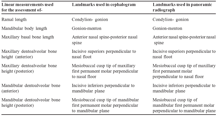
Table-I: the linear measurements measured on the lateral cephalogram and compared on the panoramic radiograp
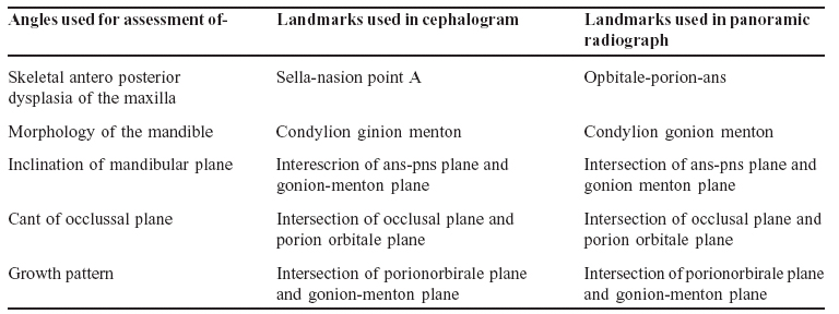
Table –II: the angular measurements measured on the lateral cephalogram and compared on the panoramic radiograph
THE LINEAR MEASUREMENTS MEASURED ON THE LATERAL CEPHALOGRAM AND
COMPARED ON THE PANORAMIC RADIOGRAPH
THE ANGULAR MEASUREMENTS MEASURED ON THE LATERAL CEPHALOGRAM AND COMPARED ON THE PANORAMIC RADIOGRAPH
RESULTS:
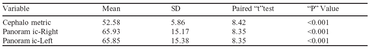
Ramal Length (CONDYLION-GONION)
Table- III (a) shows the cephalometric mean value for ramal length (condylion-gonion).
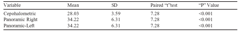
Mandibular body length (GONION-MENTON)
Maxillary Basal Bone Length
Table III (b) ( shows the cephalometric mean values of mandibular body length (gonion-mention).
Maxillary Dentoalveolar Bone Height (Anterior)
Table III (c): Shows the shows the cephalometric mean values of maxillary basel bone length (anterior nasal spineposterior nasal spine).
Maxillary Dentoalveolar Bone Height (Posterior)
Table III(d): Shows the cephalometric mean values of maxillary dentoalveolar bone height (anterior) (incisive superioris perpendicular to nasal floor) body length.
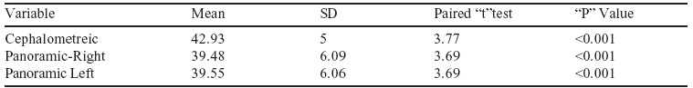
Mandibular Dentoalveolar Bone Height (Anterior)
Table III (f): Shows the cephalometric mean values of mandibular dentoalveolar bone height (anterior)(incisive
inferioris perpendicular to mandibular plane) of 42.93 mm ± 5 and panoramic mean value of 39.48 mm ± 6.09 and
39.55 mm ± 6.06 for right and left side respectively.
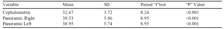
Mandibular Dentoalveolar Bone Height (Posterior)
Table III (g): Shows the cephalometric mean values of mandibular dentoalveolar bone height (Posterior)(mesiobuccal
cusp tip of mandibular first permanent molar perpendicular to mandibular plane).
Table III (g): Shows the cephalometric mean values of mandibular dentoalveolar bone height (Posterior)(mesiobuccal cusp tip of mandibular first permanent molar perpendicular to mandibular plane).
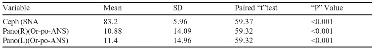
Skeletal Antero Posterior Dysplasia of the Maxilla
[Table 10]
Table IV(a): Shows the cephalometric mean value for skeletal anterior posterior dysplasis of maxilla (SNA).
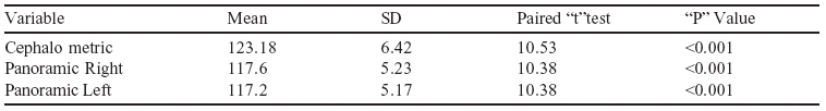
Morphology of the mandible (condylion-gonion-menton)
Table IV(b): Shows the cephalometric mean value for morphology of the mandible (condylion-gonion- menton).
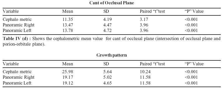
Inclination of Mandibular Plane
Table IV (c) : Shows the cephalometric mean value for inclination of mandibular plane (intersection of ANS-PNS
plane and gonion-menton plane).
Table IV (e): Shows the cephalometric mean value for growth pattern (intersection of porion orbitale plane and
gonion-menton plane).
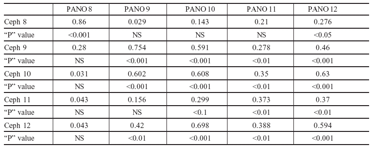
TABLE V
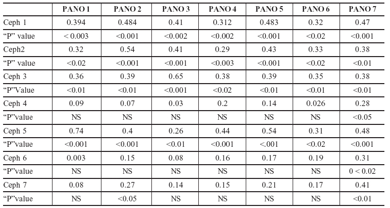
CORRELATION BETWEEN CEPHALOMETRIC AND PANORAMIC LINEAR MEASUREMENTS The values indicates Pearson’s Correlation coefficient = r
TABLE VI CORRELATION BETWEEN CEPHALOMETRIC AND PANORAMIC ANGULAR MEASUREMENTS
The value indicates Pearson’s Correlation coefficient = r
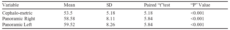
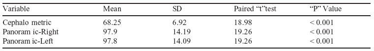
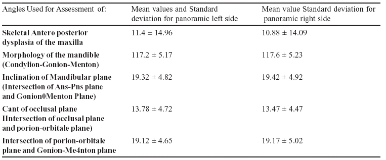
Table shows the Mean values and Standard Deviation for panoramic left and right angular parameters.
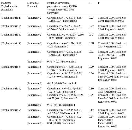
Table shows the regression equations for linear parameters in which multiple regression equation test was applied
with best possible panoramic constant to get highest predictability value.
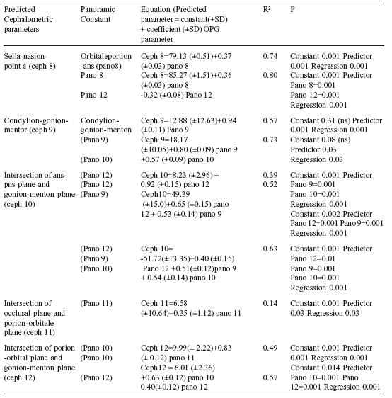
Shows the regression equations for angular parameters in which multiple regression equation test was applied
with best possible panoramic constant to get highest predictability value.
 |
 |
DISCUSSION:
Lateral cephalometry is an important tool in orthodontic diagnosis, treatment planning, and evaluation of treatment results and prediction of growth. But the major source of error in cephalometirc analysis includes radiographic film magnification, tracing, measuring, recording and landmark identification. To overcome all these limitations of lateral cephalograms, Paatero in 1961 introduced a technique called panoramic radiography which has gained a rapidly significant role in almost every field of clinical dentistry. The popularity of this technique stemmed from its simplicity of operation, its low radiation dosage when compared to conventional lateral cephalogram and full mouth radiographs, and the wide field of projected structures with reduced super imposition of investing tissue. Some limitations of lateral cephalograms may be overcome by using panoramic radiograph.
Therefore it is logical to evaluate the accuracy of measurements gleamed for panoramic radiograph as compared to lateral cephalogram for investigating dentoskeletal patterns. It is possible to measure any vertical or horizontal distance is only on one side of the mandible, either the left or the right, but that the distance must not transverse the midline of the mandible. Linear measurements are not reliable because of the magnification factor in the radiographs to eliminate the magnification factor the ratios of the linear measurements were taken and compared between lateral cephalogram and panoramic radiograph and all the radiographs were taken using same machine (PLANMECA PM-2002 EC PROLINE) and by the same operator under standard exposure conditions.
In the present study only one horizontal parameter was compared between lateral cephalogram and panoramic radiograph i.e., the maxillary basal bone length. It was found that significant amount of co-relation exist when the maxillary basal bone length was measured from lateral cephalogram and panoramic radiograph as the multiple regression equation shows 45% predictability of maxillary basal bone length from panoramic radiographs.
The weakness in radiological interpretation of linear measurements in the anterior region of the jaw contributed to investigate the reliability of vertical dimensions from panoramic radiograph. In the present study also the measurement for anterior maxillary dent alveolar heights compare between panoramic radiograph and lateral cephalogram are not reliable as evidence from the correlation coefficient value which comes out to be 0.2 (highly non significant). For posterior maxillary dent alveolar height the co-relation coefficient between panoramic value and cephalometric value is 0.54, showing that posterior maxillary dent alveolar height can be measured from panoramic radiograph. This result againfavours the study done by Xie. Q et al that vertical measurement are more reliable in the maxillary posterior region as compared to the anterior region. A mandibular anterior region measurement for panoramic radiograph and cephalogram shows the co-relation coefficient of 0.19. This study shows that predicting the cephalometric mandibular anterior dent alveolar height value from panoramic radiograph is not reliable for clinical purpose. A fourth vertical measurements used in the present study to compare were mandibular dentoalveolar height posterior. The co-relation coefficient value when compared between panoramic radiograph and cephalogram was 0.41 which is highly significant. Thus by using the anoramic mandibular dento alveolar posterior height value and panoramic ramal length value, cephalometric dentoalveolar height posterior can be predictable to significant level.In the present study, two linear oblique variables were compare i.e., ramal length (Co-Go) and mandibular body length (Go-Me) and both variable lies on one side of the mandible. The result of the present study showed that the correlation coefficient between panoramic ramal length and cephalometric ramal length was 0.394 which is clinical significant. For mandibular body length (Go-Me), the correlation coefficient between panoramic value and cephalometric value in the present study is 0.54 which is highly significant. Various studies have been done in the past to investigate the reliability of angular measurements taken from panoramic radiograph. The values for the gonial angle are of particular interest because lateral caphalograms do not permit reliable registration of this angle and the super imposition images create difficulties in recognition and measurement of the individual angle. This disadvantage is not encountered in panoramic radiography, which has proved to be as accurate as cephalography in determining the gonial angle. The co-relation between cephalometric and panoramic radiograph measurements found that gonial angles of panoramic radiograph and basal plane angles of cephalogram showed high correlation of 0.49. Thus the present study is in favour with all the previous studies that gonial angle can be predicted reliably from the panoramic radiograph. The predictability of cephalometric mandibular plane angles from panoramic radiograph have been studied and concluded that instead of panoramic mandibular plane angles giving highest predictability value for mandibular plane angles,
it was panoramic basal plane angle whose correlation coefficient with cephalometric mandibular plane angle was remarkably high i.e., upto 0.76. The present study also favours the study done by Ackam et al. Hence it is recommended that, to predict the cephalometric mandibular plane angle, both panoramic mandibular plane and panoramic basal plane angle should be used.
The present study used panoramic angle (orbitaleporion-ANS) to predict cephalometric SNA angle and found that correlation coefficient was 0.86, slightly less than what Ackam deduced in his study. The regression equation also shows that cephalometric SNA angle can be predicted from panoramic (orbitale-porion-ANS) angle by 74% predictability. Basal plane angle between lateral cephalometric and panoramic radiographs was also compared in this study and the correlation coefficient was 0.608 suggesting that cephalometric basal plane angles can be predicted to significant level from panoramic radiograph.
But the regression equation shows that the predictability level using panoramic gonial angle, basal plane angle and mandibular plane angle to predict cephalometric basal plane angle increases upto 63%.
It was found that the correlation coefficient between cephalometric cant of occlusal plane and panoramic cant of occlusal plane was just 0.373, suggesting that predicting cephalometric cant of occlusal plane from panoramic radiograph is nor reliable. The regression equation also shows that the predictability level was just 14%. Thus an orthodontist should be cautious while comparing cephalometric cant of occlusal plane from panoramic radiograph. No significance difference was found between
angular measurements for panoramic left and right side in the present study and all the angular and linear measurements were not influenced by the sex of an individual.
SUMMARY AND CONCLUSION:
The present study was undertaken in an attempt to answer whether panoramic radiograph can be used instead of lateral cephalogram to assess dentoskeletal pattern of the patient.
THE RESULT OF THIS STUDY SHOWED THAT :
1. All angular parameters measured on panoramic radiograph showed high correlation and predictability when compared
with similar parameters measured on lateral cephalogram especially for gonial angle measurements except for cant of occlusal
plane, which shows least predictability when measured from panoramic radiograph. Thus panoramic radiographs can be used for angular measurements instead of lateralce phalogram.
2. For vertical parameters measured on panoramic radiographs, in posterior region (molar area) the correlation and predictability was acceptable and clinically significant when compared with similar parameters in lateral cephalogram, but the measurements done in the maxillary and mandibular anterior region were not reliable when measured from a panoramic radiograph. Thus a clinician should be careful while measuring vertical measurements in maxillomandibular anterior region from panoramic radiographs.
3. Oblique and horizontal measurements done on panoramic radiograph also shows that these measurements can be recorded from panoramic radiograph with high predictability.
4. No significance difference was found between angular measurements for panoramic left and right side in the present study and all the angular and linear measurements were not influenced by the sex of an individual.
REFERENCES:
Broadbent B.H.: A new x-ray technique and its application to orthodontia. Angle Orthod 1931; 1 : 45-66.
Bjork A., Skieller V.: Normal and abnormal growth of the mandible. A synthesis of longitudinal cephalometirc implant
studies over a period of 25 years. European Journal of Orthodontics 1983; 5: 1-46.
Graber T.M.: Panoramic radiography in orthodontic diagnosis. American Journal of Orthodontics. 1967; 53 (111): 799-821.
Paatero Y.V.: Pantomography and Orthopantomography. Oral Surg, Oral Med and Oral Path.1961; 14: 947-953.
Coaz P.W., White S.C.: Oral Radiology: Principles and interpretation, 1987; 2nd Edition.
Hauck R.M.: Documentation of tooth movement by means of panoral radiograph. Am J Orthod 1970: 57(4): 386-392.
Philipp R.G., Hurst R.V.: The cant of the occlusal plane and distortion in the panoramic radiograph. Angle Orthod 1978;
48: 317-32.
Lucchesi M.V., Wood R.E., et al: Suitability of the panoramic radiograph for assessment of mesiodistal angulation
of teeth in the buccal segments of the mandible. Am J Orthod Dentofac Orthop 1988; 94: 303-10.
Mckee I.W., Williamson P.C., et al: The accuracy of 4 panoramic units in the projection of mesiodistal tooth angulations. Am J Orthod Dentofacial Orthop 2002; 121: 166-75.
Mckee I.W., Glover K.E., et. al: The effect of vertical and horizontal head positioning in panoramic radiography on mesiodistal tooth angulations. Angle Orthod 2001; 71: 442- 451.
Stramotas S., Geenty J.P., et. al: Accuracy of linear and angular
measurements on panoramic radiographs taken at various
positions in vitro. European Journal of Orthodontics 2002;
24: 43-52.
Slagsvold O., Pederson K.: Gonial Angle Distortion In Lateral had Films: A Methodological Study. Am J Orthod 1977; 71: 554-564.
Mattila K., Altoren M. and Haavikka K.: Determination of the gonial angle from the orthopantogram. Angle Orthodontists.
1997; 47: 107-110.
Ursi W.J.S., Almeida R.R., et. al: Assessment of mesiodital axial inclination through panoramic radiography. Journal of
Clinical Orthodontics 1990; 24 (3): 166-173. Latheim T.A., Svanaes D.B.: Reproducibility of rotational
panoramic radiography: Mandibular linear dimensions and angles. Am J Orthod Dentofacial Orthop 1986; 90: 45-51.
Samawi S.S.B., Burke P.H.: Angular distortion in the orhtopantomogram. British Journal of Orthodontics 1984; 11: 100-107.
Rejebian G.P.: A statistical correlation of individual tooth size distortions on the orthopantomographic radiograph. Am J Orthod 1979; 75 (5): 524-534.
Thanyakarn C., Hansen K., et. al: Measurements of tooth length in panoramic radiographs. 2: Observer performance. Dentomaxillofac Radiol 1995; 21: 31-35. |