INTRODUCTION
Knowledge of the internal dental morphology is a complex and extremely important point for planning and performing of endodontic therapy. The several anatomical variations existing in the root canal system may contribute for failure of
root canal therapy, mainly in teeth with pulp necrosis.
Maxillary molars have been reported to have extra canals in as much as 95% of the times (13). It has been suggested that using a No. 1 round bur or ultrasonic instruments to remove secondary dentin from the pulpal floor along the
mesiobuccal-palatal aspect of the molar triangle will uncover an additional 31% of these orifices.(8) An earlier study found these secondary canals 69% of the time in vitro but only 31% in vivo (9). Another in vivo study found two canals in the mesiobuccal roots of maxillary first molars 77% of the time, and, of these, 62% had two apical foramina (10). A fourth root in maxillary molars is reported to be rare (0.4%). (11, 12) This paper reports the case of a maxillary left first molar that presented three root canals in the mesiobuccal root. Root canal therapy and case management are described.
CASE REPORT
A 15-year old male patient reported to the deptt of pedodontics, Himachal Dental College Sundernagar for treatment of tooth # 26. The patient was complaining of occasional pain in the tooth and was sealed with a zinc oxide and eugenol filling. Thermal sensitivity tests were negative indicating non vital pulp. Neither any fistulae nor any edema was present. The periapical radiograph showed a small area of thickened periodontal ligament around the root apices. (Fig 1)
Local anesthetic (2%Lidocaine with 1:200000 epinephrine) was given and an access opening was made. The operative field was thereafter isolated with a rubber dam. Exploration of root canal entrances was done with an endodontic explorer. The exploration revealed 3 distinct canals in the mesiobuccal root, 1 canal in the distobuccal root and 1 canal in the palatal root, which were later further confirmed by the radiograph for working length determination (Fig 2).
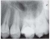 | Fig 1: Initial Radiograph
 |
All 5 canals were explored with #10 K-files (Mani Japan). After exploring the canals with #10 K-files, #15 K-files (Dentsply) were introduced in the mesiobuccal-1, mesiobuccal-2 and mesiopalatal canals, while #20 and #25 K-files
(Dentsply) were introduced in the distobuccal and palatal canals, respectively, to determine the working length. The preparation of the canals was done with Protaper files (Dentsply). Preparation of the mesiobuccal canal-1, mesiobuccal canal-2 and mesiopalatal canals was done till file F1 size. Apical preparation of the distobuccal and palatal canals was performed till size F1 and F2 respectively. The canals were repeatedly irrigated with sodium hypochlorite during the preparation.
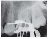 | Fig 2: Determination of working length
|
Chemomechanical preparation was completed in the first session. The canals were flushed with saline in the end, dried and filled with calcium hydroxide paste, which was used as an intracanal medication. Root canal access was sealed with a zinc oxide and eugenol dressing. After 7 days, the canals were emptied, copiously irrigated with 2% sodium hypohlorite and then with saline and dried with paper points. Main gutta-percha cones were selected for each canal and all canals were filled using the lateral condensation technique. A final radiograph was taken to confirm the ompleteness and extension of root filings. The tooth was provisionally sealed and the patient was recalled later for restorative treatment.
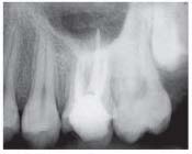 | Fig 2: Determination of working length
|
DISCUSSION
The variations in dental anatomy play an important role in root canal therapy. A great predominance of two very close canals in the mesiobuccal root of maxillary molars has been demonstrated (2). Despite the current high success rate achieved in endodontic treatments, the mesiobuccal root is still associated to a considerable number of failures due to the difficulty in locating and filling the second and/ or third mesiobuccal canals (1,2). On account of this, root canal therapy of these teeth should be carried out using angulated x-rays (4,5),efficient explorers, wider crown accesses (5), adequate lighting and, whenever possible, image agnification (6,7). In the case reported in this paper, the mesiobuccal root presented a moderate
curvature with three canals. The mesiobuccal canal-1 had one opening and one exit, while the mesiobuccal canal-2 and the mesiopalatal canal presented two openings and one exit. The instrumentation of these canals was carried out with nickel-titanium files. These instruments are indicated in these cases due to their flexibility and because they pose lesser risks of
step formation or perforations. The instrumentation technique (crown down) used in this study recommend a wide access to the middle and cervical thirds, which facilitated the cleaning of the apical third and the filling of the root canals (3).
CONCLUSION
Treating teeth with multiple canals is a fairly common problem. It is a fact that makes imperative a careful search in every tooth for additional canals.
REFERENCES
1. Vertucci FJ. Root canal anatomy of the human permanent teeth. Oral Surg Oral Med Oral Pathol 1984;58:589-599.
2. Fogel HM, Christie WH, Peikoff MD. Canal configuration in the mesiobuccal root of the maxillary first molar: a clinical study. J Endod 1994;20:135-137.
3. Siqueira JF Jr, Rocas IN, Santos SR, Lima KC, Magalhães FA, De Uzeda M. Efficacy of instrumentation techniques and
irrigation regimens in reducing the bacterial population within root canals. J Endod 2002;28:181-184.
4. Weine FS, Eskoz N. Canal configuration of the mesiobuccal root of the maxillary second molar. J Endod 1995;21:38-42.
5. Weller RN, Hartwell GR. The impact of improved access and searching techniques on detection the mesiolingual canal in
maxillary molars. J Endod 1989;15:82-83.
6. Kim S. Principles of endodontic microsurgery. Dent Clin North Am 1997;41:481-497.
7. Kim S, Baek S. The microscope and endodontics. Dent Clin North Am 2004;48:11-18.
8. Kulild JC, Peters DD. Incidence and configuration of canal systems in the mesiobuccal root of maxillary first and second
molars. JOE 1990;16:311.
9. Bjorndal AM, Skidmore AE. Anatomy and morphology of human teeth. Iowa City (IA): Univ. of Iowa; 1983.
10. Neaverth EJ, et al. Clinical investigation (in vivo) of endodontically treated maxillary first molars. JOE 1987;10:506.
11. Fahid A. Maxillary second molar with three buccal roots. JOE 1988;14:181.
12. Libfeld H, Rolstein I. Incidence of four-rooted maxillary second molars: Literature review and radiographic survey of 1200
teeth. JOE 1989;15:129.
13. Ingle JI, Bakland LK. Endodontics 5th Ed, 2002; 10:416 |