INTRODUCTION
Salivary gland tumors accounts for less than 3% of head and neck tumors.They are more common in adults than in children.1 Among all the salivary gland tumors, pleomorphic adenoma is most commonly encountered lesion accounting
for approximately 60% of salivary gland tumors. Most salivary gland tumors occur in major salivary glands, especially in parotid gland. As for as intraoral salivary gland tumors are concerned, pleomorphic adenoma also ranks most frequently
encountered lesion. Palate is most commonly affected site followed by upper lip and buccal mucosa respectively.2
CASE REPORT
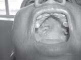
A 45-year-old female patient visited the department of oral medicine and radiology with c/o swelling in right maxillary posterior region which had duration of one year. History revealed that patient noticed this swelling one year back and is gradually increasing in size. Swelling was painless and not associated with any ulceration and discharge. There was no preceding history of trauma and her past medical history was unremarkable. The general health of the patient is preserved. On examination a 1.5x2 cm sized, firm, non-tender, circumscribed lesion in the relation to 16, 17, 18 region of hard palate was observed. Fig (1). It was adherent to the underlying structures and overlying mucosa was intact and pink in color. There was no regional lymphadenopathy and her nasal examination was within normal limits. General physical and systemic examination was also normal.
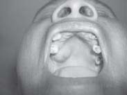 | Fig 1 – Pleomorphic Adenoma in right posterior region of hard palate
 |
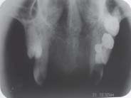 | Fig 2 - Radiograph showing no bony invasion
|
A clinical differential diagnosis of odontogenic cyst or minor salivary gland tumor, was considered. An incisional biopsy revealed abenign tumor having characteristic features of pleomorphic adenoma on histopathologic examination. Radiograph of maxilla (occlual view) did not show bony invasion. Figure (2). The entire tumor was excised with a wide
margin and the underlying bone was drilled out. Fig (3) and fig (4). There has been no recurrence for 6 months during follow up. DISCUSSION Pleomorphic adenomas are usually painless, slow-growing tumors; however, some cases
exhibiting rapid growth have been reported.3,4,5. The term pleomorphic describes the embryogenic basis of origin of these tumors, which contains both epithelial and mesenchymal tissues.6 It has been postulated that these tumors arise from intercalated and myoepithelial cells. Most cases of palatal pleomorphic adenoma cause only a bulge in the palatal mucosa, but some cause an erosion of the palatal bone as well.3, 7. These adenomas usually present as asymptomatic submucosal swellings although a few cases have exhibited ulceration and bleeding, usually resulting from trauma. The tumor presents morphologically diverse features, however, both epithelial and mesenchymal elements must be present for diagnosis.1, 2 Histopathology reveals a tumor composed of islands of stellate and spindle cells that are interspersed in a myxoid background. The pleomorphic nature is determined by an inner layer of epithelial cells and an outer layer of myoephithelial cells arranged in a variety of patterns associated with scant or
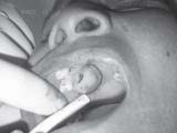 | Fig 3 Tumor outline demarcated
|
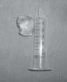 | Fig 4 Excised Mass
|
 | Fig 5 showing post operative healing
|
abundant stroma. Variation may include squamous metaplasia, calcification, cartilage-like tissue, oxyphillic cells and rarely malignant transformation.7 Plain x-ray and hematologic investigations play no part in the diagnosis of salivary gland tumor of the palate.8 The noninvasive diagnostic aids for salivary gland tumors include ultrasound, CT, and magnetic resonance imaging (MRI). These are useful methods in determining the size of the lesion as well as verifying any bony involvement. CT and MRI both provide important information on the location, size, and extension of the tumor into the surrounding superficial and deep tissues. CT is superior to MRI in evaluating bone, especially in diagnosing erosion and perforation of the bony palate and possible involvement of the nasal cavity or maxillary sinus.8,9 MRI, with its high resolution for soft tissue, provides better definition of the vertical and inferior tumor extension through its multiplanar
capacity and the tumor–muscle interface9 and more clearly indicates the degree of encapsulation.8Another advantage of MRI over CT is the absence of exposure to radiation and intravenous contract medium. Treatment of palatal pleomorphic adenoma involves wide local excision of the tumor by careful dissection of the palatal mucosa from the encapsulated mass, including its surrounding capsule, together with clear margins involving the periosteum and associated mucosa, followed by curettage of the underlying bone with a sharp spoon or bur under copious sterile normal saline irrigation, to avoid recurrence.3, 4 These tumors usually do not recur after adequate surgical excision. Most recurrences can be attributable to inadequate surgical techniques such as simple enucleation leaving behind microscopic pseudopod-like extensions.
REFERENCES
1. LunaMA, BatasakisJG, EL-NaggarAK, Salivary gland tumorsin children, Ann Otol Rhinol Laryngol.1991; 100:869-71.
2. Kittipongdhanuthai, Kraisory sappayatosok, Kosak kongin, Buccal Pleomorphic adenoma of palate in child. A case report. Med oral Patol oral cir 2009; 100:869-71.
3. De Courten, T. Lombardi and J. Samson, Pleomorphic adenoma in a child: 9-year follow-up, Int J Oral Maxillofac
Surg 1996; 25: 293.
4. J.L. Lopez-Cedrum, G. Gonzalez-Landa and B. Birichinaga,Pleomorphic adenoma of the palate in children: Report of a
case, Int J Oral Maxillofac Surg 1996; 25:206.
5. H. Shabaan, J. Bruce and P.J. Davenport, Recurrent pleomorphic adenoma of the palate in a child, Br J Plast Surg
2001; 54: 245.
6. Batsakis JG. Neoplasms of the minor and ‘lesser’ major salivary glands. In: Tumors of the Head and Neck. The
Williams and Wilkins Company, Baltimore. 1981; 38-47.
7. W.H. Crawford and L.H. Guernsey, Pleomorphic adenoma of the palate: Report of a case, Oral Surg Oral Med Oral
Pathol 1967; 23: 116.
8. M.A. Pogrel, The management of salivary gland tumors of the palate, J Oral Maxillofac Surg 1994; 52: 454.
9. A. Noghreyan, A. Gatot and E. Maor et al., Palatal pleomorphic adenoma in a child, J Laryngol Otol 1995; 109:343. |